6 Respiratory System
Learning Objectives
- Identify general terms related to the respiratory system
- Spell the respiratory system medical terms and use correct abbreviations
- Identify the medical specialties associated with the respiratory system
- Recognize common diseases, disorders, and procedures related to the respiratory system
Chapter Six: Table of Contents
What is it? – General Terms: The Respiratory System
What Can Go Wrong? – Diseases, Disorders, and Conditions
- Nose and Pharynx
- Larynx
- Bronchial Tree
- Lungs
- Pleura and Thorax
- Signs and Symptoms of Respiratory Disorders
How Do We Fix it or Make it Better?
References, Attributions, and Image Descriptions
Respiratory System Word Parts
General Terms
| Term | Word Breakdown | Description |
|---|---|---|
| respiratory (rEs-puhr-ruh-tor-ee) |
-atory pertaining to re- spir/o |
The respiratory system is the network of organs and tissues that help you breathe. It includes your airways, lungs and blood vessels. Read More |
| respiration (res-puhr-rAY-shuhn) |
-ation process; condition re- spir/o |
breathing in and breathing out |
| inhalation (in-huh-lAY-shuhn) |
-ation process; condition in- hal/o |
Breathing in |
| inspiration (in-spuhr-rAY-shuhn) |
-ation process; condition in- spir/o |
Breathing in |
| exhalation (eks-huh-lAY-shuhn) |
-ation process; condition ex- hal/o |
Breathing out |
| expiration(ek-spuhr-rAY-shun) | -ation process; condition ex- spir/o |
Breathing out |
Click on prefixes, combining forms, and suffixes to reveal a list of word parts to memorize for the Respiratory System.
Respiratory Basics
Introduction to the Respiratory System
How long you can hold your breath as you continue reading… How long can you do it? Chances are you are feeling uncomfortable already. A typical human cannot survive without breathing for more than three minutes, and even if you wanted to hold your breath longer, your autonomic nervous system would take control. Although oxygen is critical for cells, it is the accumulation of carbon dioxide that primarily drives your need to breathe.
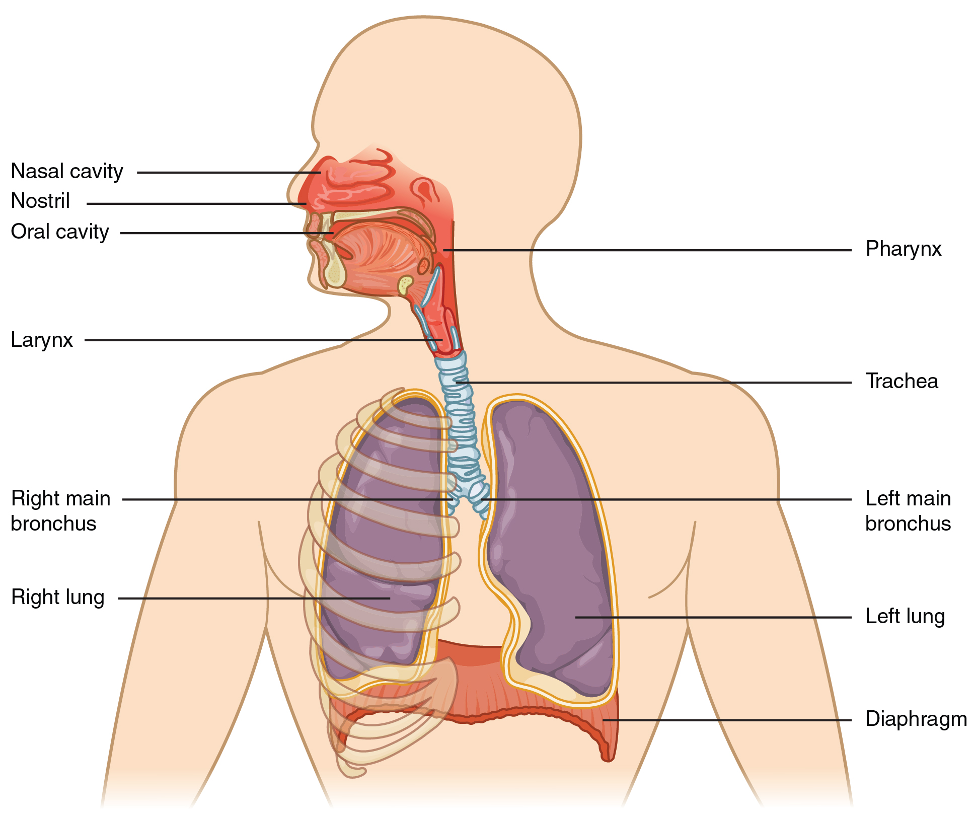
The major structures of the respiratory system function primarily to provide oxygen to body tissues for cellular respiration, remove the waste product carbon dioxide, and help to maintain acid-base balance. Portions of the respiratory system are also used for non-vital functions, such as sensing odors, speech production, and for straining, such as coughing.
Watch this video:
Media 7.1. Respiratory System: From Inspiration to Expiration Explained in Simple Words [Online video]. Copyright 2015 by Science ABC.
Respiratory System Medical Terms
Anatomy (Structures) of the Respiratory System
The Nose and its Adjacent Structures
The major entrance and exit for the respiratory system is through the nose. When discussing the nose, it is helpful to divide it into two major sections:
The nares open into the nasal cavity, which is separated into left and right sections by the nasal septum (Figure 7.2). The nasal septum is formed anteriorly by a portion of the septal cartilage and posteriorly by the perpendicular plate of the ethmoid bone and the thin vomer bones.
Each lateral wall of the nasal cavity has three bony projections the inferior conchae are separate bones and the superior and middle conchae are portions of the ethmoid bone. Conchae increase the surface area of the nasal cavity, disrupt the flow of air as it enters the nose, causing air to bounce along the epithelium, where it is cleaned and warmed. The conchae and meatuses trap water during exhalation preventing dehydration.
The floor of the nasal cavity is composed of the hard palate and the soft palate. Air exits the nasal cavities via the internal nares and moves into the pharynx.
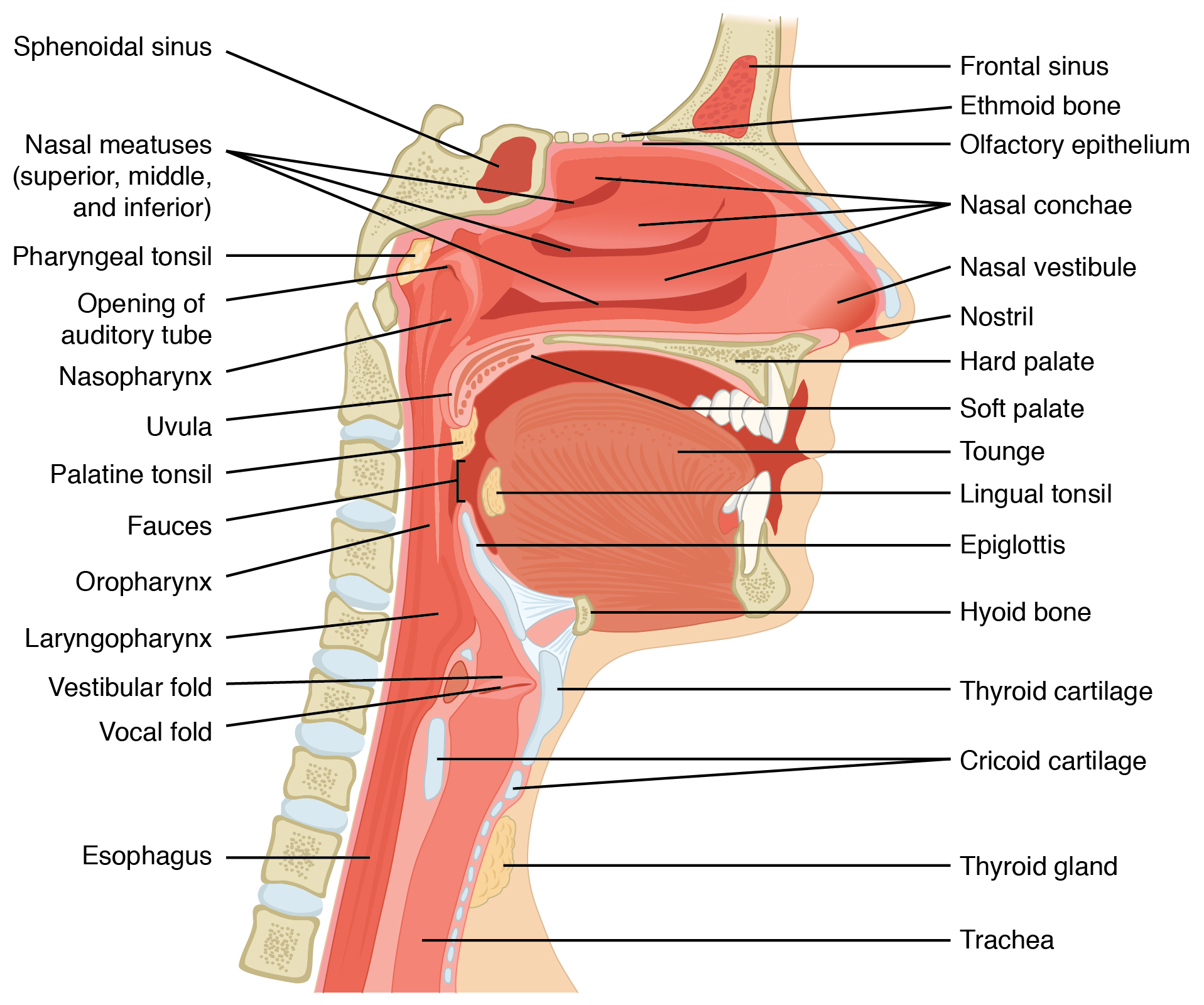
Paranasal sinuses, serve to warm and humidify incoming air and are lined with a mucosa which produces mucus. Paranasal sinuses are named for their associated bone:
- frontal sinus
- maxillary sinus
- sphenoidal sinus
- ethmoidal sinus
The nares and anterior portion of the nasal cavities are lined with mucous membranes, containing sebaceous glands and hair follicles that serve to prevent the passage of large debris, such as dirt, through the nasal cavity. An olfactory epithelium used to detect odors is found deeper in the nasal cavity.
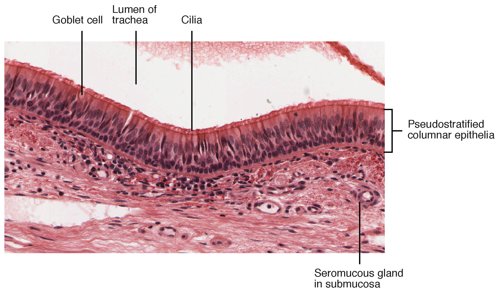
The conchae, meatuses, and paranasal sinuses are lined by respiratory epithelium composed of pseudostratified ciliated columnar epithelium (Figure 7.3). The epithelium contains specialized epithelial cells that produce mucus to trap debris. The cilia of the respiratory epithelium help to remove mucus and debris with a constant beating motion, sweeping materials towards the throat to be swallowed.
This moist epithelium functions to warm and humidify incoming air. Capillaries located just beneath the nasal epithelium warm the air by convection. Serous and mucus-producing cells also secrete defensins, immune cells patrol the connective tissue providing additional protection.
Pharynx
The pharynx is divided into three major regions: the nasopharynx, the oropharynx, and the laryngopharynx (see Figure 7.4).
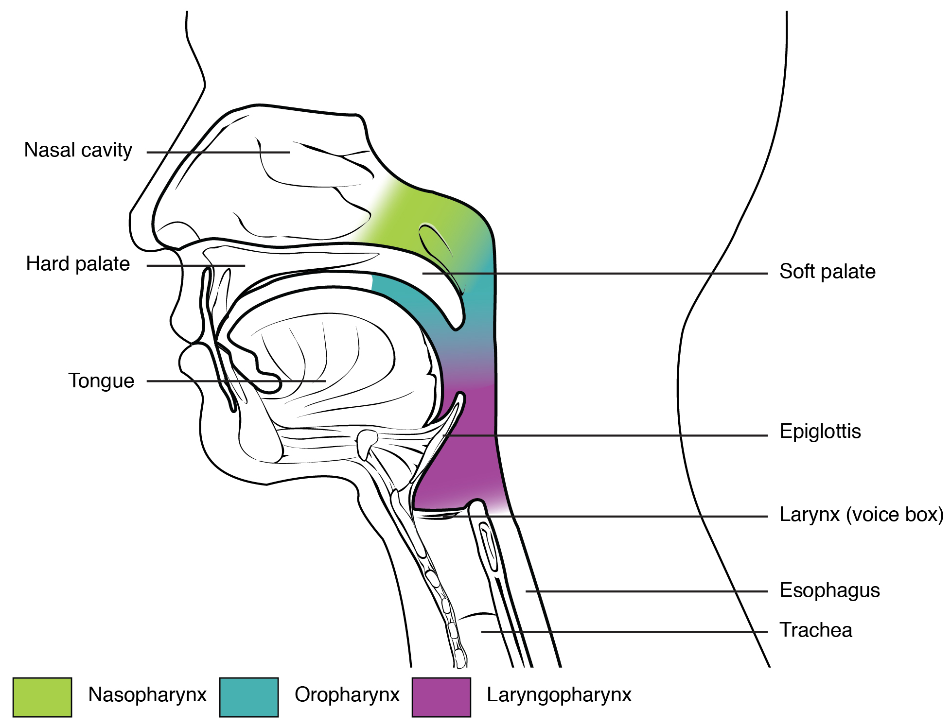
At the top of the nasopharynx are the pharyngeal tonsils. The function of the pharyngeal tonsil is not well understood, but it contains a rich supply of lymphocytes and is covered with ciliated epithelium that traps and destroys invading pathogens that enter during inhalation. The pharyngeal tonsils are large in children, but tend to regress with age and may even disappear. The uvula and soft palate move like a pendulum during swallowing, swinging upward to close off the nasopharynx to prevent ingested materials from entering the nasal cavity. Auditory (Eustachian) tubes that connect to each middle ear cavity open into the nasopharynx. This connection is why colds often lead to ear infections.
The oropharynx is bordered superiorly by the nasopharynx and anteriorly by the oral cavity. The oropharynx contains two distinct sets of tonsils:
- The palatine tonsils.
- A palatine tonsil is one of a pair of structures located laterally in the oropharynx in the area of the fauces.
- The lingual tonsils.
- The lingual tonsil is located at the base of the tongue.
Similar to the pharyngeal tonsil, the palatine and lingual tonsils are composed of lymphoid tissue, and trap and destroy pathogens entering the body through the oral or nasal cavities.
The laryngopharynx is inferior to the oropharynx and posterior to the larynx. It continues the route for ingested material and air until its inferior end, where the digestive and respiratory systems diverge. The stratified squamous epithelium of the oropharynx is continuous with the laryngopharynx. Anteriorly, the laryngopharynx opens into the larynx, whereas posteriorly, it enters the esophagus.
Larynx
The structure of the larynx is formed by several pieces of cartilage. Three large cartilage pieces form the major structure of the larynx.
- Thyroid cartilage (anterior):
- The thyroid cartilage is the largest piece of cartilage that makes up the larynx. The thyroid cartilage consists of the laryngeal prominence, or “Adam’s apple,” which tends to be more prominent in males.
- Epiglottis (superior):
- Three smaller, paired cartilages—the arytenoids, corniculates, and cuneiforms—attach to the epiglottis and the vocal cords and muscle that help move the vocal cords to produce speech.
- Cricoid cartilage (inferior):
- The thick cricoid cartilage forms a ring, with a wide posterior region and a thinner anterior region.
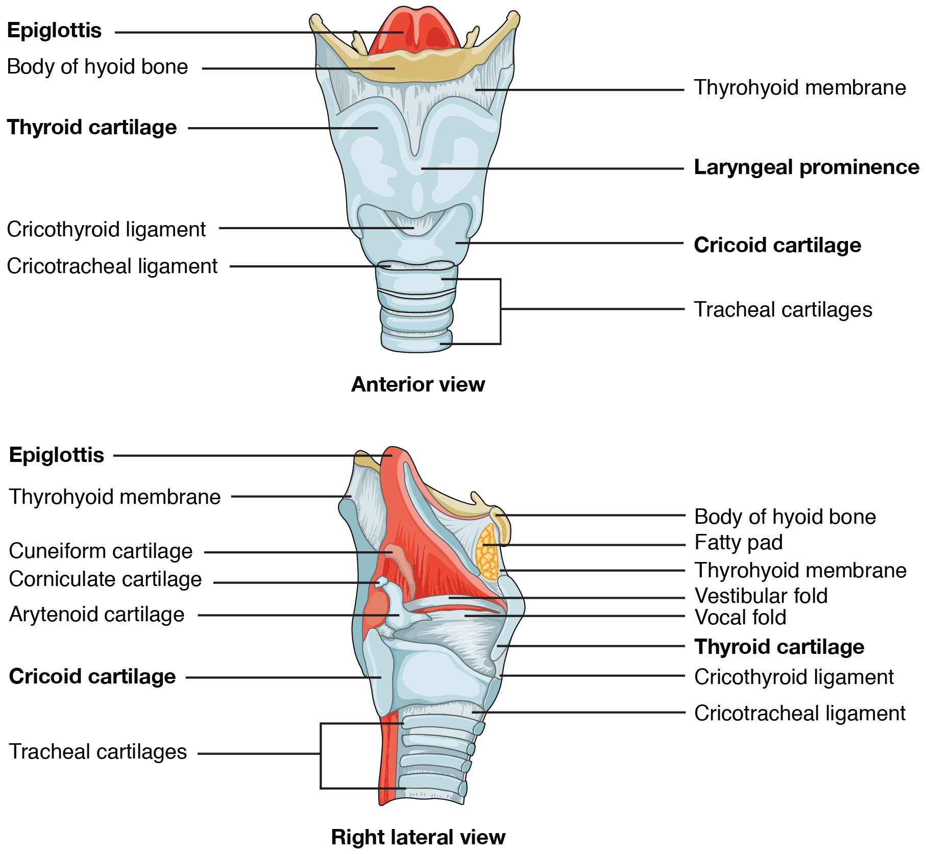
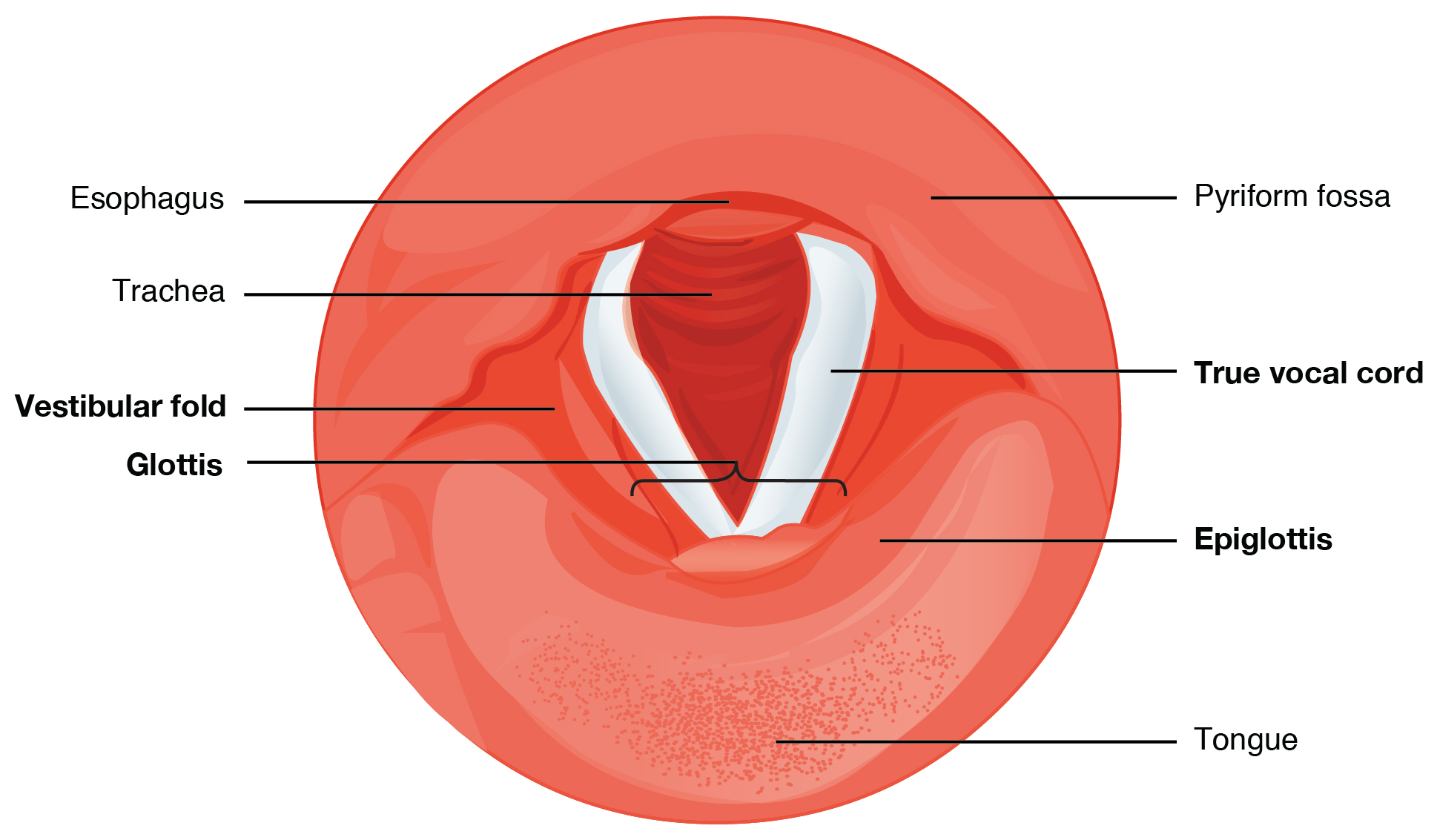
When the epiglottis is in the “closed” position, the unattached end of the epiglottis rests on the glottis. A vestibular fold, or false vocal cord, is one of a pair of folded sections of mucous membrane. A true vocal cord is one of the white, membranous folds attached by muscle to the thyroid and arytenoid cartilages of the larynx on their outer edges. The inner edges of the true vocal cords are free, allowing oscillation to produce sound.
The act of swallowing causes the pharynx and larynx to lift upward, allowing the pharynx to expand and the epiglottis of the larynx to swing downward, closing the opening to the trachea. These movements produce a larger area for food to pass through, while preventing food and beverages from entering the trachea.
Similar to the nasal cavity and nasopharynx, this specialized epithelium produces mucus to trap debris and pathogens as they enter the trachea. The cilia beat the mucus upward towards the laryngopharynx, where it can be swallowed down the esophagus.
Trachea
The trachea is formed by 16 to 20 stacked, C-shaped pieces of hyaline cartilage that are connected by dense connective tissue. The trachealis muscle and elastic connective tissue together form the fibroelastic membrane. The fibroelastic membrane allows the trachea to stretch and expand slightly during inhalation and exhalation, whereas the rings of cartilage provide structural support and prevent the trachea from collapsing. The trachealis muscle can be contracted to force air through the trachea during exhalation. The trachea is lined with pseudostratified ciliated columnar epithelium, which is continuous with the larynx. The esophagus borders the trachea posteriorly.
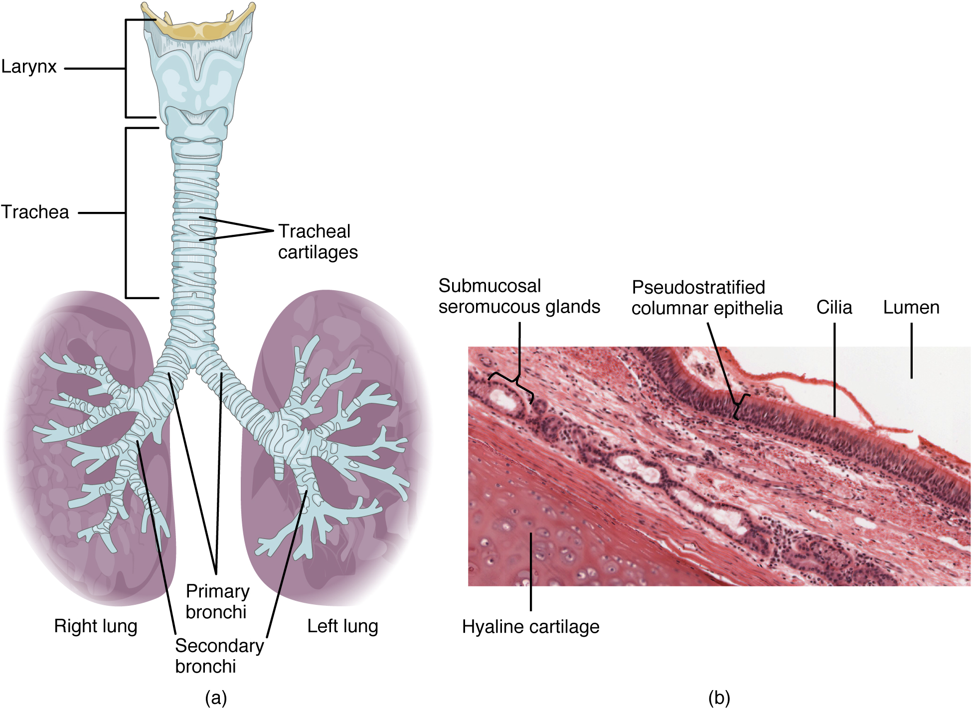
Bronchial Tree
The trachea branches into the right and left primary bronchi at the carina. These bronchi are also lined by pseudostratified ciliated columnar epithelium containing mucus-producing goblet cells (Figure 7.7b). The carina is a raised structure that contains specialized nervous tissue that induces violent coughing if a foreign body, such as food, is present. Rings of cartilage, similar to those of the trachea, support the structure of the bronchi and prevent their collapse. The primary bronchi enter the lungs at the hilum. The bronchi continue to branch into bronchial a tree. A bronchial tree (or respiratory tree) is the collective term used for these multiple-branched bronchi. The main function of the bronchi, like other conducting zone structures, is to provide a passageway for air to move into and out of each lung. The mucous membrane traps debris and pathogens.
A bronchiole branches from the tertiary bronchi. Bronchioles, which are about 1 mm in diameter, further branch until they become the tiny terminal bronchioles, which lead to the structures of gas exchange. There are more than 1000 terminal bronchioles in each lung. The muscular walls of the bronchioles do not contain cartilage like those of the bronchi. This muscular wall can change the size of the tubing to increase or decrease airflow through the tube.
Respiratory Zone
In contrast to the conducting zone, the respiratory zone includes structures that are directly involved in gas exchange. The respiratory zone begins where the terminal bronchioles join a respiratory bronchiole, the smallest type of bronchiole (see Figure 7.8), which then leads to an alveolar duct, opening into a cluster of alveoli.
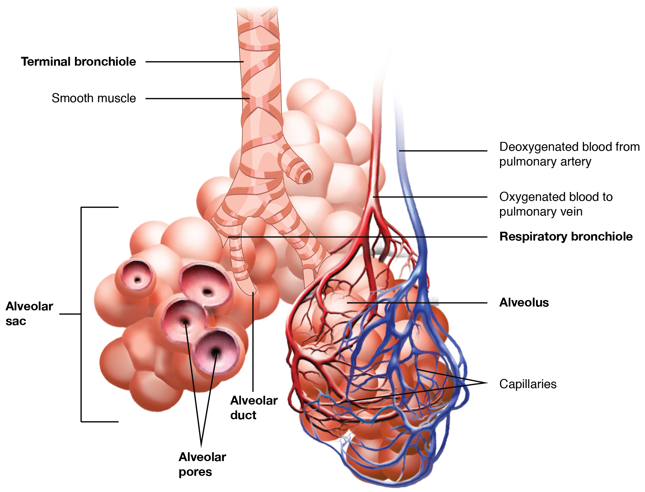
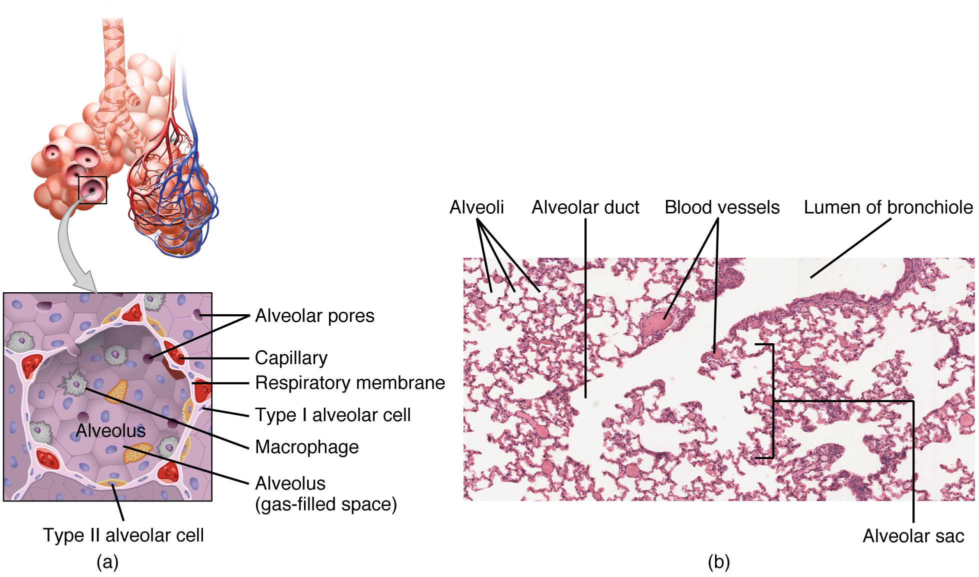
Alveoli
An alveolar duc opens into a cluster of alveoli. An alveolus is one of the many small, grape-like sacs that are attached to the alveolar ducts. An alveolar sac is a cluster of many individual alveoli that are responsible for gas exchange. An alveolus is approximately 200 μm in diameter with elastic walls that allow the alveolus to stretch during air intake, which greatly increases the surface area available for gas exchange. Alveoli are connected to their neighbors by alveolar pores, which help maintain equal air pressure throughout the alveoli and lung (see Figure 7.9).
Concept Check
- What are the components of the bronchial tree?
- What is the purpose of cilia?
- Where does gas exchange take place?
Gross Anatomy of the Lungs
The lungs are pyramid-shaped, paired organs that are connected to the trachea by the right and left bronchi; on the inferior surface, the lungs are bordered by the diaphragm. The lungs are enclosed by the pleurae, which are attached to the mediastinum. The right lung is shorter and wider than the left lung, and the left lung occupies a smaller volume than the right. The cardiac notch allows space for the heart (see Figure 7.10). The apex of the lung is the superior region, whereas the base is the opposite region near the diaphragm. The costal surface of the lung borders the ribs. The mediastinal surface faces the mid line.

Each lung is composed of smaller units called lobes. Fissures separate these lobes from each other. The right lung consists of three lobes: the superior, middle, and inferior lobes. The left lung consists of two lobes: the superior and inferior lobes. A pulmonary lobule is a subdivision formed as the bronchi branch into bronchioles. Each lobule receives its own large bronchiole that has multiple branches. An interlobular septum is a wall, composed of connective tissue, which separates lobules from one another.
Can you correctly label the respiratory system structures?
Physiology (Function) of the Respiratory System
Blood Supply
The major function of the lungs is to perform gas exchange, which requires blood from the pulmonary circulation.
- This blood supply contains deoxygenated blood and travels to the lungs where erythrocytes pick up oxygen to be transported to tissues throughout the body.
- The pulmonary artery carries deoxygenated, arterial blood to the alveoli.
- The pulmonary artery branches multiple times as it follows the bronchi, and each branch becomes progressively smaller in diameter.
- One arteriole and an accompanying venule supply and drain one pulmonary lobule. As they near the alveoli, the pulmonary arteries become the pulmonary capillary network.
- The pulmonary capillary network consists of tiny vessels with very thin walls that lack smooth muscle fibers.
- The capillaries branch and follow the bronchioles and structure of the alveoli. It is at this point that the capillary wall meets the alveolar wall, creating the respiratory membrane.
- Once the blood is oxygenated, it drains from the alveoli by way of multiple pulmonary veins, which exit the lungs through the hilum.
Nervous Innervation
The blood supply of the lungs plays an important role in gas exchange and serves as a transport system for gases throughout the body. Innervation by the both the parasympathetic and sympathetic nervous systems provides an important level of control through dilation and constriction of the airway.
- The parasympathetic system causes bronchoconstriction.
- The sympathetic nervous system stimulates bronchodilation.
Reflexes such as coughing, and the ability of the lungs to regulate oxygen and carbon dioxide levels, also result from autonomic nervous system control. Sensory nerve fibers arise from the vagus nerve, and from the second to fifth thoracic ganglia. The pulmonary plexus is a region on the lung root formed by the entrance of the nerves at the hilum. The nerves then follow the bronchi in the lungs and branch to innervate muscle fibers, glands, and blood vessels.
Pleura of the Lungs
Each lung is enclosed within a cavity that is surrounded by the pleura. The pleura (plural = pleurae) is a serous membrane that surrounds the lung. The right and left pleurae, which enclose the right and left lungs, respectively, are separated by the mediastinum.
The pleurae consist of two layers:
- The visceral pleura is the layer that is superficial to the lungs, and extends into and lines the lung fissures (see Figure 7.11).
- The parietal pleura is the outer layer that connects to the thoracic wall, the mediastinum, and the diaphragm.
The visceral and parietal pleurae connect to each other at the hilum. The pleural cavity is the space between the visceral and parietal layers.
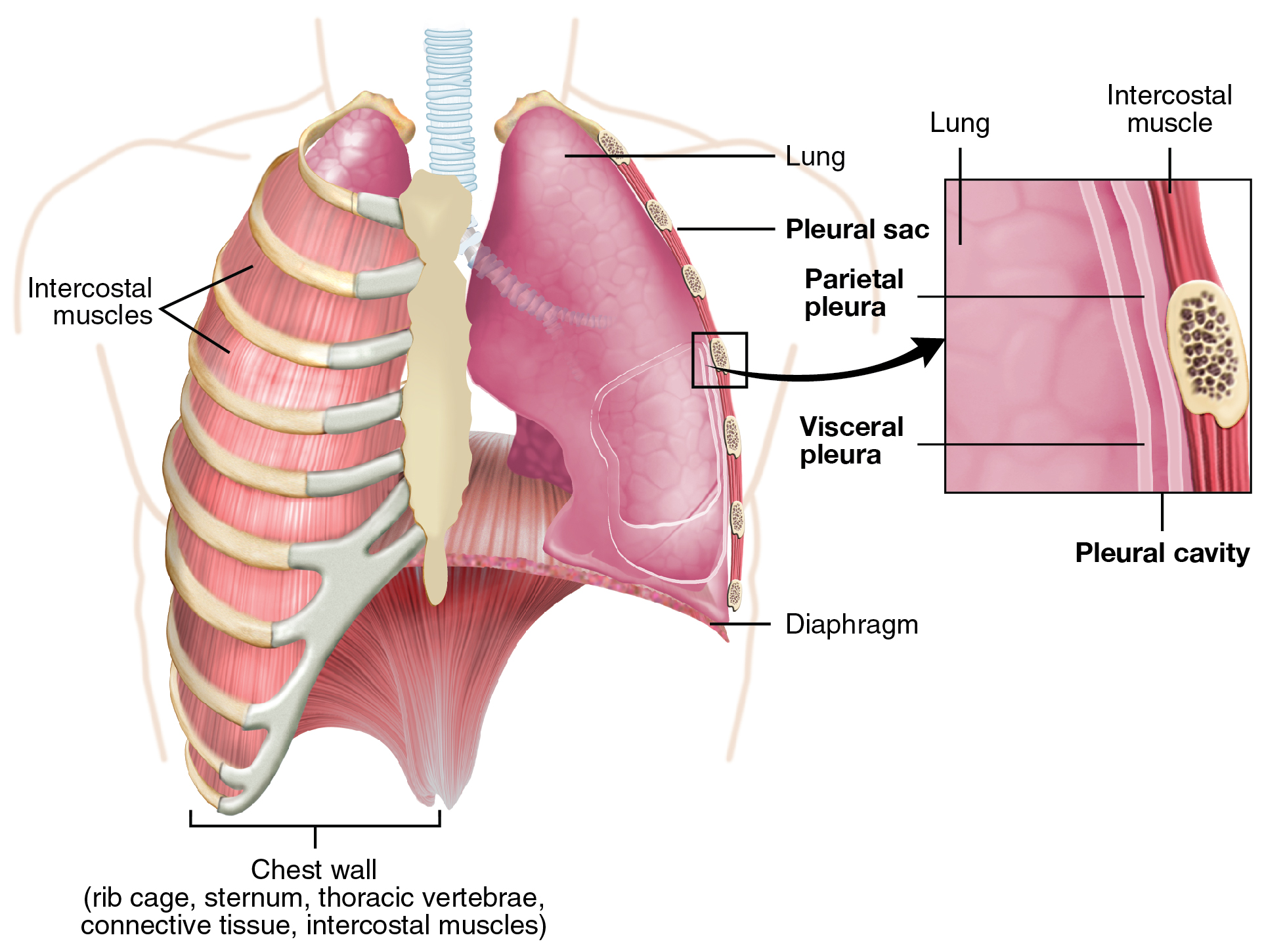
The pleurae perform two major functions:
- Produce pleural fluid that that lubricates surfaces, reduces friction to prevent trauma during breathing, and creates surface tension that helps maintain the position of the lungs against the thoracic wall. This adhesive characteristic of the pleural fluid causes the lungs to enlarge when the thoracic wall expands during ventilation, allowing the lungs to fill with air.
- The pleurae also create a division between major organs that prevents interference due to the movement of the organs, while preventing the spread of infection.
Pulmonary Ventilation
The difference in pressures drives pulmonary ventilation because air flows down a pressure gradient, that is, air flows from an area of higher pressure to an area of lower pressure.
- Air flows into the lungs largely due to a difference in pressure; atmospheric pressure is greater than intra-alveolar pressure, and intra-alveolar pressure is greater than intrapleural pressure.
- Air flows out of the lungs during expiration based on the same principle; pressure within the lungs becomes greater than the atmospheric pressure.
Pulmonary ventilation comprises two major steps: inspiration and expiration. Inspiration is the and expiration (Figure 7.12). A respiratory cycle is one sequence of inspiration and expiration.
Two muscle groups are used during normal inspiration the diaphragm and the external intercostal muscles. Additional muscles can be used if a bigger breath is required.
- The diaphragm contracts, it moves inferiorly toward the abdominal cavity, creating a larger thoracic cavity and more space for the lungs.
- The external intercostal muscles contract and moves the ribs upward and outward, causing the rib cage to expand, which increases the volume of the thoracic cavity.
Due to the adhesive force of the pleural fluid, the expansion of the thoracic cavity forces the lungs to stretch and expand as well. This increase in volume leads to a decrease in intra-alveolar pressure, creating a pressure lower than atmospheric pressure. As a result, a pressure gradient is created that drives air into the lungs.
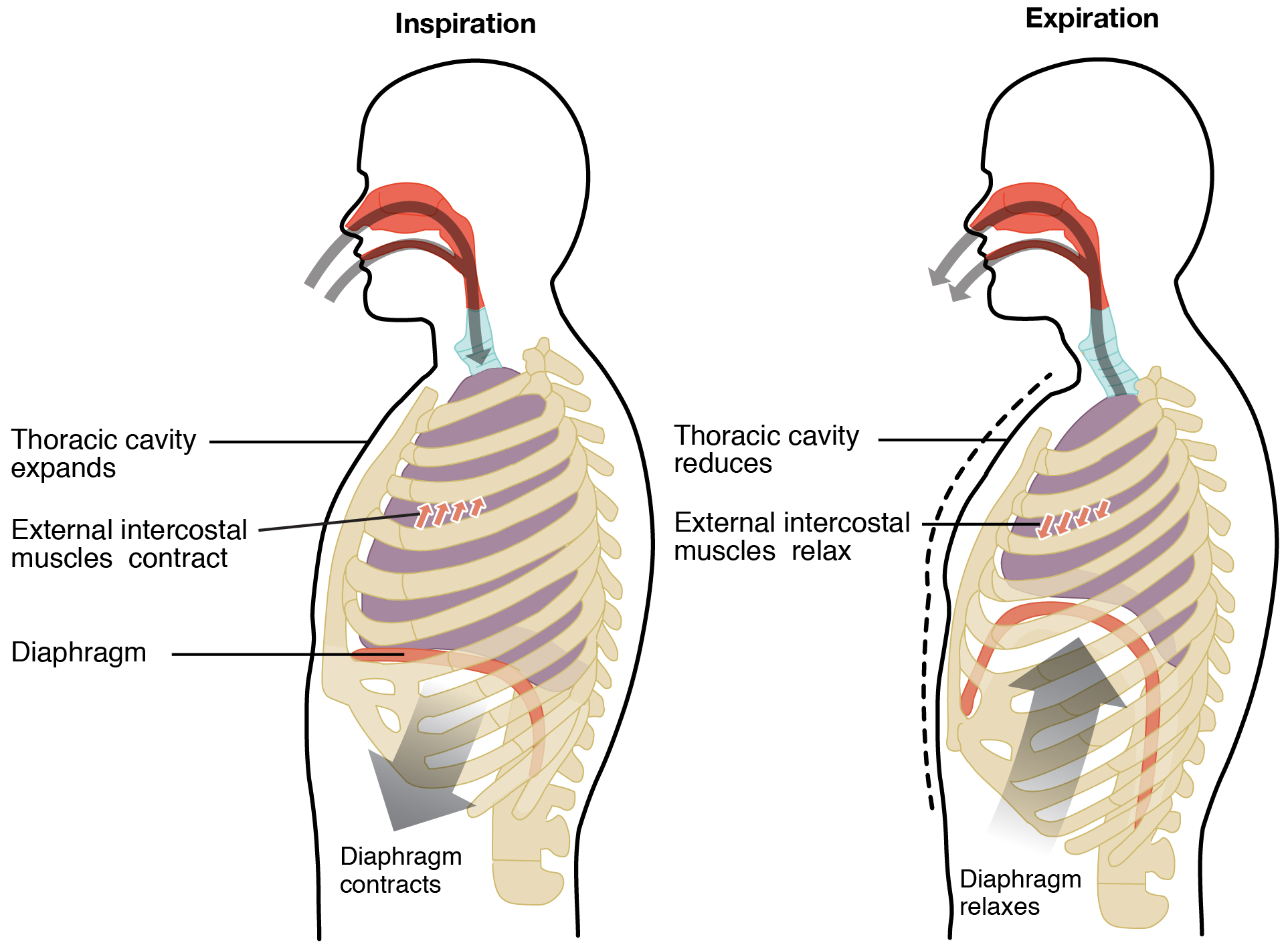
The process of normal expiration is passive, meaning that energy is not required to push air out of the lungs.
- The elasticity of the lung tissue causes the lung to recoil, as the diaphragm and intercostal muscles relax following inspiration.
- The thoracic cavity and lungs decrease in volume, causing an increase in interpulmonary pressure. The interpulmonary pressure rises above atmospheric pressure, creating a pressure gradient that causes air to leave the lungs.
There are different types, or modes, of breathing that require a slightly different process to allow inspiration and expiration:
- Quiet breathing, also known as eupnea, is a mode of breathing that occurs at rest and does not require the cognitive thought of the individual. During quiet breathing, the diaphragm and external intercostals must contract.
- Diaphragmatic breathing, also known as deep breathing, requires the diaphragm to contract. As the diaphragm relaxes, air passively leaves the lungs.
- Costal breathing, also known as a shallow breath, requires contraction of the intercostal muscles. As the intercostal muscles relax, air passively leaves the lungs.
- Forced breathing, also known as hyperpnea, is a mode of breathing that can occur during exercise or actions that require the active manipulation of breathing, such as singing.
- During forced breathing, inspiration and expiration both occur due to muscle contractions. In addition to the contraction of the diaphragm and intercostal muscles, other accessory muscles must also contract.
- During forced inspiration, muscles of the neck contract and lift the thoracic wall, increasing lung volume.
- During forced expiration, accessory muscles of the abdomen contract, forcing abdominal organs upward against the diaphragm. This helps to push the diaphragm further into the thorax, pushing more air out. In addition, accessory muscles help to compress the rib cage, which also reduces the volume of the thoracic cavity.
- During forced breathing, inspiration and expiration both occur due to muscle contractions. In addition to the contraction of the diaphragm and intercostal muscles, other accessory muscles must also contract.
Concept Check
- Breathing normally, place your hand on your stomach take in one full respiratory cycle.
- What type of breathing are you doing?
- Keeping your hand on your stomach, take in one large breath and exhale.
- What type of breathing are you doing?
- Complete 10 jumping jacks, once completed, place your hand on your stomach and take in one full respiratory cycle.
- What type of breathing are you doing?
Respiratory Rate and Control of Ventilation
Breathing usually occurs without thought, although at times you can consciously control it, such as when you swim under water, sing a song, or blow bubbles. The respiratory rate is the total number of breaths that occur each minute. Respiratory rate can be an important indicator of disease, as the rate may increase or decrease during an illness or in a disease condition. The respiratory rate is controlled by the respiratory center located within the medulla oblongata in the brain, which responds primarily to changes in carbon dioxide, oxygen, and pH levels in the blood.
The normal respiratory rate of a child decreases from birth to adolescence:
- A child under 1 year of age has a normal respiratory rate between 30 and 60 breaths per minute.
- By the time a child is about 10 years old, the normal rate is closer to 18 to 30.
- By adolescence, the normal respiratory rate is similar to that of adults, 12 to 18 breaths per minute.
Watch this video:
Media 7.1. Respiratory System, Part 2: Crash Course A&P #32 [Online video]. Copyright 2015 by CrashCourse.
Medical Terms not Easily Broken into Word Parts
Diseases, Disorders, and Conditions
Diseases, Disorders, and Conditions of the Nose and Pharynx
| Term | Word Breakdown | Description |
|---|---|---|
| anosmia (a-nAHz-mee-uh) |
-ia condition an- osm/e |
Absence of the sense of smell |
| epistaxis (ep-uh-stAk-suhs) |
Nasal hemorrhage or nosebleed | |
| pharyngitis (far-uhn-jIE-tuhs) |
-itis inflammation pharyng/o |
Inflammation of the pharynx or sore throat |
| rhinitis (rie-nIE-tuhs) |
-itis inflammation rhin/o |
Rhinitis is when a reaction occurs that causes a stuffy nose, runny nose, sneezing, and itching. . Read more about types and symptoms |
| sinusitis (sie-nyuh-sIE-tuhs) |
-itis inflammation sinus/o |
Sinusitis is an inflammation of the tissues in your sinuses (spaces in your forehead, cheeks and nose usually filled with air). It causes facial pain, a stuffy or runny nose, and sometimes a fever and other symptoms. It’s usually caused by the common cold, but other viruses, bacteria, fungi and allergies can also cause sinusitis. Read more |
| tonsillitis (tahn-suh-lIE-tis) |
-itis inflammation tonsil/o |
Tonsillitis is an infection of your tonsils, two masses of tissue at the back of your throat. Read more |
| upper respiratory infection (URI) | The upper respiratory system includes the nose, mouth, pharynx, and larynx. An upper respiratory infection affects the upper part of your respiratory system, including your sinuses and throat. Upper respiratory infection symptoms include a runny nose, sore throat and cough (from post-nasal drip). It is also called a common cold. Read more |
Nose in the Pharynx of time!
Diseases, Disorders, and Conditions of the Larynx
| Term | Word Breakdown | Description |
|---|---|---|
| laryngitis (lair-uhn-jIE-tuhs) |
-itis inflammation laryng/o |
Inflammation of the larynx. Typical symptoms include hoarseness and loss of voice. |
| stridor (strIE-duhr) |
An abnormal high-pitched sound you make when you inhale or exhale. This sound happens when you have a blockage in your throat (pharynx), voice box (larynx) or windpipe (trachea). Read more |
Larynx Knowledge!
Diseases, Disorders, and Conditions of the Bronchial Tree
| Term | Word Breakdown | Description |
|---|---|---|
| asthma (Az-muh) |
A condition in which your airways narrow and swell and may produce extra mucus. This can make breathing difficult and trigger coughing, a whistling sound (wheezing) when you breathe out and shortness of breath. Read more | |
| bronchiectasis (brahng-kee-Ek-tuh-suhs) |
-ectasis dilation; dilatation; widening bronch/o |
A chronic condition where the walls of the bronchi are thickened from inflammation and infection. People with bronchiectasis have periodic flare-ups of breathing difficulties, called exacerbations. Read more |
| bronchitibronchitiss (brahn-kIE-tuhs) |
-itis inflammation bronch/o |
An inflammation of the lining of your bronchial tubes, which carry air to and from your lungs. People who have bronchitis often cough up thickened mucus, which can be discolored. Bronchitis may be either acute or chronic. Read more |
Asthma
Asthma is a common chronic condition that affects all age groups. In 2011/2012 there were 3.8 million Canadians diagnosed with asthma and a disproportionate number of children and youth (Government of Canada, 2018). To learn more, visit the Asthma in Canada Data Blog . Asthma is a chronic disease characterized by inflammation, edema of the airway, and bronchospasms which can inhibit air from entering the lungs. Bronchospasms can lead to an “asthma attack.” An attack may be triggered by environmental factors such as dust, pollen, pet hair, or dander, changes in the weather, mold, tobacco smoke, and respiratory infections, or by exercise and stress (Betts, et al., 2013).
Symptoms of an asthma attack involve coughing, shortness of breath, wheezing, and tightness of the chest. Symptoms of a severe asthma attack require immediate medical attention and may include dyspnea that results in cyanotic lips or face, confusion, drowsiness, a rapid pulse, sweating, and severe anxiety. The severity of the condition, frequency of attacks, and identified triggers influence the type of medication that an individual may require. Longer-term treatments are used for those with more severe asthma. Short-term, fast-acting drugs that are used to treat an asthma attack are typically administered via an inhaler. For young children or individuals who have difficulty using an inhaler, asthma medications can be administered via a nebulizer (Betts, et al., 2013.
Diseases, Disorders, and Conditions of the Lungs
| Term | Word Breakdown | Description |
|---|---|---|
| atelectasis (at-uh-lEk-tuh-suhs) |
-ectasis expansion; dilation atel/o |
When your airways or the tiny sacs at the end of them don’t expand the way they should when you breathe. Also known as a collapsed lung. Read more |
| chronic obstructive pulmonary disease (COPD) (pUl-muh-nair-ee) |
-ic Pertaining to chron/o |
COPD is a term used to represent a number of persistant, long -term respiratory diseases including chronic bronchitis and emphysema. Video that explains COPD |
| cystic fibrosis (CF) (sIs-tik fie-brOH-suhs) |
-ic pertaining to cyst/o -osis fibr/o |
A progressive, genetic disease that affects the lungs, pancreas, and other organs. Over time, it becomes harder to breathe and increases the risk of lung infections Read more and watch videos |
| emphysema (em-fuh-zEE-muh) |
-ectasis dilation; dilatation; widening bronch/o |
A pathologic accumulation of air in tissues or organs. Emphysema develops over time and involves the gradual damage of lung tissue, specifically the destruction of the alveoli (tiny air sacs). Gradually, this damage causes the air sacs to rupture and create one big air pocket instead of many small ones. This reduction in the lung surface area traps air in the damaged tissue and prevents oxygen from moving through the bloodstream. Additionally, this blockage causes the lungs to slowly overfill and makes breathing increasingly more difficult and is characterized by a persistent cough. Read more Video that explains emphysema |
| empyema (em-pie-EE-muh) |
-ema condition em- py/o |
A collection of pus in the pleural cavity. The pleural cavity surrounds each of the lungs. |
| hypercapnia (hie-puhr-kAp-nee-uh) |
-ia condition hyper- capn/o |
Excess carbon dioxide in the blood. Read more |
| influenza (in-floo-En-zuh) |
A contagious respiratory illness caused by influenza viruses that infects the nose, throat, and sometimes the lungs. Symptoms include sore throat, fever, cough, and body aches . It can cause mild to severe illness, and at times can lead to death. Read more | |
| lower respiratory infection | A type of lower respiratory infection that affects one or both lungs. It causes the air sacs, or alveoli, of the lungs to fill up with fluid or pus. Bacteria, viruses, or fungi may cause pneumonia. Video that explains pneumonia | |
| pneumoconiosis (noo-moh-koh-nee-OH-suhs) |
-osis condition pneumon/o- coni/o |
A group of lung diseases caused by the lung’s reaction inhaling certain dusts. Also known as occupational pneumonia Read more |
| pneumonia (nu-mOH-nyuh) |
-ia condition pneumon/o- |
A type of lower respiratory infection that affects one or both lungs. It causes the air sacs, or alveoli, of the lungs to fill up with fluid or pus. Bacteria, viruses, or fungi may cause pneumonia. Video that explains pneumonia |
| pulmonary edema (pUl-muh-nair-ee) (i-dEE-muh) |
-ary condition pulmon/o- |
Pulmonary is a term that means pertaining to the lungs. Edema means swelling or abnormally large amounts of fluid in the intercellular tissue spaces of the body, Pulmonary edema is too much fluid in the lungs. Pulmonary edema is different than pneumonia as pneumonia comes from an infection and pulmonary edema comes from other causes such as heart conditions. |
| pulmonary embolism (pUl-muh-nair-ee) (Em-buh-liz-uhm) |
-ary condition pulmon/o- -ism embol/o- |
Pulmonary is a term that means pertaining to the lungs. Embolism is the sudden blocking of an artery by a clot or foreign material in the blood. A pulmonary embolism (PE) is a blockage of the pulmonary arteries in the lung that scan cause a lack of blood flow leading to lung tissue damage. It can cause low blood oxygen levels that can damage other organs in the body, too. Read more |
| tuberculosis (TB) (tu-buhr-kyuh-lOH-suhs) |
An infectious disease caused by tubercles on the lungs. Infectious diseases are illnesses caused by germs (such as bacteria, viruses, and fungi) that enter the body, multiply, and can cause an infection. Tuberculosis is caused by a different bacteria than pneumonia. Symptoms of TB are coughing, slow fever, and tiredness. Pneumonia does not only effect the lungs. TB can also affect the urinary and skeletal system. Video that explains TB |
Chronic Obstructive Pulmonary Disease (COPD)
COPD is a term used to represent a number of respiratory diseases including chronic bronchitis and emphysema. COPD is a chronic condition with most symptoms appearing in people in their middle 50s. Symptoms include shortness of breath, cough, and sputum production. Symptoms during flare ups or times of exacerbation, may include green or brown mucous, increase in the viscosity or amount of mucus, chest pain, fever, swollen ankles, headaches, dizziness, and blue lips or fingers There is no cure for COPD. Shortness of breath may be controlled with bronchodilators. The best plan is to avoid triggers and getting sick. Clients with COPD are advised to avoid people who are sick, get the flu shot and reduce their exposure to pollution and cigarette smoke. While there are several risk factors 80% of cases are associated with cigarette smoking (Government of Canada, 2018). To learn more about COPD visit the Public Health Agency of Canada’s web page on COPD.
Lung Cancer
Lung cancer is a leading cause of cancer death among both males and females in Canada with 98% occurring in adults over 50. Symptoms often appear in the late stages with 50% being diagnosed at STAGE IV (Government of Canada, 2019a). Symptoms may include shortness of breath, wheezing, blood in the mucus, chronic chest infections, dysphagia, pleural effusion, and enlarged lymph nodes. There are two types of lung cancer, small cell lung cancer (SCLC) linked to cigarette smoking, grows quickly and metastasizes. Non-small cell lung cancer (NSCLC) is more common and grows slowly. Changes in lung cells may lead to benign tumours or malignant tumours. Cancers that start in other parts of the body may metastasize to the lungs. Risk factors include smoking, air pollution, family history exposure to second-hand smoke, exposure to radon gas, and exposure to carcinogens (Government of Canada, 2019). Treatment will depend on the type of lung cancer and the stage at diagnosis. Treatments may include surgery, chemotherapy, targeted therapy, immunotherapy, and radiation therapy (Government of Canada, 2019a).
Breath Taking Knowledge!
The Effects of Second-Hand Tobacco Smoke
- It is estimated that the risk of developing lung cancer is increased by up to 30 percent in nonsmokers who live with an individual who smokes in the house, as compared to nonsmokers who are not regularly exposed to second-hand smoke.
- Children who live with an individual who smokes inside the home have a larger number of lower respiratory infections, which are associated with hospitalizations, and higher risk of sudden infant death syndrome (SIDS). Second-hand smoke in the home has also been linked to a greater number of ear infections in children, as well as worsening symptoms of asthma (Betts, et al., 2013).
Diseases, Disorders, and Conditions of the Pleura and Thorax
| Term | Word Breakdown | Description |
|---|---|---|
| hemothorax | -thorax chest hem/o |
When blood collects in the chest; usually caused by injury. |
| pleural effusion plUR-uhl i-fyOO-zhuhn |
-ation process; condition re- spir/o |
Effusion means escaping fluid.
Pleural effusion is an excessive accumulation of fluid in the pleural space. |
| pleurisy plUR-uh-see |
Inflammation of the sheet-like layers that cover the lungs (the pleura). The most common symptom of pleurisy is a sharp chest pain when breathing deeply. Read more | |
| pneumothorax noo-muh-thOR-aks |
-thorax chest pneum/o |
A type of collapsed lung. Pneumothorax happens when air leaks into the pleural cavity. This is the space between the outside of your lung and your ribcage. Excess air in the pleural cavity (often occurs with a hemothorax). This is different from an atelectasis which is caused by a blockage of the air passages |
Practice Makes Perfect!
Signs and Symptoms of Breathing Problems
| Term | Word Breakdown | Description |
|---|---|---|
| anoxia (a-nAHk-see-uh) |
-ia condition an- ox/o |
A total lack of oxygen; |
| apnea Ap-nee-uh |
-pnea breathing Prefix: |
Temporary cessation of breathing, especially during sleep. |
| asphyxia as-fIk-see-uh |
Not enough oxygen in your body. Causes include allergic reactions, drowning and foreign objects blocking your airway. Symptoms include trouble breathing, loss of consciousness and inability to speak. You can prevent asphyxiation by being cautious. Treatment includes CPR, the Heimlich maneuver and medications. Read more | |
| bradypnea | -pnea breathing brady- |
Slow breathing |
| cyanosis sie-uh-nOH-suhs |
-osis condition cyan/o- |
A bluish discoloration of the skin resulting from poor circulation or inadequate oxygenation of the blood. |
| dyspnea dIs-nee-uh |
-pnea breathing dys- |
Difficult breathing |
| expectoration ik-spek-tuhr-rAY-shuhn |
-ation process; condition ex- |
The act of coughing up and spitting out materials from the respiratory tract. |
| eupnea yoop-nEE-uh |
-pnea breathing eu- |
Easy or normal respiration. |
| hemoptysis hi-mAHp-tuh-suhs |
-ptysis spitting hem/o eu- |
Spitting up blood |
| hyperpnea hie-puhr-nEE-uh |
-pnea breathing hyper- |
Abnormal increase in the depth of breathing. May or may not have an increased rate of breathing. |
| hypoxemia hi-pahk-sEE-mee-uh |
-emia blood condition hypo- ox/o |
Deficient oxygenation of the blood; |
| hypoxia hi-pAHk-see-uh |
-ia condition hypo- |
A deficiency of oxygen reaching the tissues of the body. Video that Video that explains the difference between hypoxia and hypoxemia |
| orthopnea | -pnea breathing orth/o- |
Difficulty in breathing that occurs when lying down and is relieved upon changing to an upright position |
| paroxysmal nocturnal dyspnea (PND) par-uhk-sIz-muhl nahk-tUHR-nl) (dIs-nee-uh |
A sudden attack of shortness of breath that occurs at night | |
| purulent | Consisting of, containing, or discharging pus. | |
| tachypnea | -pnea breathing tachy- |
Increased respiratory rate |
Sleep Apnea
Sleep apnea is a chronic disorder that occurs in children and adults. It is characterized by the cessation of breathing during sleep. These episodes may last for several seconds or several minutes, and may differ in the frequency with which they are experienced. Sleep apnea leads to poor sleep, symptoms include fatigue, evening napping, irritability, memory problems, morning headaches, and excessive snoring. A diagnosis of sleep apnea is usually done during a sleep study, where the patient is monitored in a sleep laboratory for several nights. Treatment of sleep apnea commonly includes the use of a device called a continuous positive airway pressure (CPAP) machine during sleep. The CPAP machine has a mask that covers the nose, or the nose and mouth, and forces air into the airway at regular intervals. This pressurized air can help to gently force the airway to remain open, allowing more normal ventilation to occur (Betts, et al., 2013).
Respiratory System: Medical and Surgical Procedures
| Term | Word Breakdown | Description |
|---|---|---|
| auscultation and percussion aw-skuhl-tAY-shuhn puhr-kUHsh-uhn |
Auscultation is listening to the internal sounds of the body, usually using a stethoscope. Percussion involves tapping the body to elicit sounds and determining whether the sounds are appropriate for a particular organ or area of the body. | |
| bronchoscopy brahn-kAH-skuh-pee |
-scopy examine: for examining |
A procedure that shows the lungs and air passages. During bronchoscopy, a thin tube (bronchoscope) is passed through the nose or mouth, down the throat and into the lungs. Video that explains bronchoscopy |
| cardiopulmonary resuscitation (CPR) kahr-dee-oh-pUl-muh-nair-ee ri-suhs-uh-tAY-shuhn |
A medical procedure involving repeated compression of a patient's chest, performed in an attempt to restore the blood circulation and breathing of a person who has suffered cardiac arrest. | |
| incentive spirometer (IS) in-sEn-tiv spie-rAH-muh-tuhr |
Device used to exercise the lungs. Directions to use an incentive spirometer |
|
| lobectomy loh-bEk-tuh-mee |
-ectomy to surgical cut out lob/o |
Surgical removal of a lobe, such as a lobe of the lung |
| oximetry ahk-sIm-uh-tree |
-metry process of measuring Combining form: |
The use of an oximeter to measure oxygen saturation rates. The measurement reflects the saturation of your red blood cells with oxygen. Normal rates are 95 to 100%. |
| pneumonectomy noo-muh-nEk-tuh-mee |
-ectomy to surgical cut out pneumon/o |
To surgically remove of an entire lung or of one or more lobes of a lung |
| thoracentesis tho·ra·cen·te·sis |
-centesis surgical puncture to remove fluid thorac/o |
Removal of fluid from the chest. A needle is inserted between the ribs in the back and fluid is withdrawn. Read more |
| thoracotomy thor-uh-kAH-tuh-mee |
-tomy to cut into thorac/o |
A surgical procedure in which a cut is made between the ribs to see and reach the lungs or other organs in the chest or thorax. A thoracotomy is performed for diagnosis or treatment of a disease and allows doctors to visualize, biopsy or remove tissue as needed. American Lung Association
|
| tracheostomy tray-kee-AH-stuh-mee |
-ostomy to surgically create a new opening trache/o |
A hole that is made through the front of the neck and into the windpipe (trachea). A tracheostomy tube is placed into the hole to keep it open for breathing. A tracheostomy provides an air passage to help you breathe when the usual route for breathing is somehow blocked or reduced. Read more |
Dissecting the Procedures
Respiratory System: Drug Categories
| Term | Word Breakdown | Description |
|---|---|---|
| antitussive an-tee-tUHs-iv |
-ive pertaining to anti- tuss/i |
A cough suppressant |
| bronchodilators brahng-koh-die-lAY-tuhrz |
-or one who dilay/o bronch/o |
A drug that relaxes bronchial muscle resulting in expansion of the bronchial air passages |
| expectorant ik-spEk-truhnt |
-ant doing; having ex- pector/o |
An agent that promotes the discharge or expulsion of mucus from the respiratory tract by thinning and loosening mucus in the respiratory tract. |
Respiration Drugs
Medical Specialties: Careers Related to the Respiratory System
| Term | Word Breakdown | Description |
|---|---|---|
| respiratory therapist rEs-puhr-ruh-tor-ee thAIR-uh-pist |
A respiratory therapist (RT) is trained to help people with lung diseases or disorders that can result from a myriad of issues. They assess and treat patients dealing with pulmonary distress due to complications from asthma, bronchitis, COPD, pneumonia, chest trauma, prematurity, lung cancer, and more. Respiratory therapists are in high-demand, too, with industry growth projected to be at least 23 percent over the next decade according to the U.S. Bureau of Labor Statistics.Video that describes respiratory therapy | |
| pulmonologist pul-muh-nAH-luh-juhst |
-ist specialist pulmon/o log/o |
.A pulmonologist is a doctor who diagnoses and treats diseases of the respiratory system -- the lungs and other organs that help you breathe.
For some relatively short-lasting illnesses that affect your lungs, like the flu or pneumonia, you might be able to get all the care you need from your regular doctor. But if your cough, shortness of breath, or other symptoms don't get better, you might need to see a pulmonologist |
Respiratory Therapists (RTs)
Respiratory Therapists (RTs) are health care professionals that monitor, assess and treat people who are having problems breathing. RTs are regulated which means they must be a member of the College of Respiratory Therapists of Ontario to work as an RT in Ontario. RTs are trained in ventilation and airway management, cardiopulmonary resuscitation, oxygen and aerosol therapy. They care for patients during cardiac stress-testing, pulmonary function testing, smoking cessation, high-risk births, rehabilitation, and surgery. They treat patients with asthma, bronchitis, COPD, emphysema, heart disease, and pneumonia (College of Respiratory Therapists of Ontario, n.d.). For more information, visit the College of Respiratory Therapist’s What is a Respiratory Therapist? web page.
Thoracic Surgeon
A thoracic surgeon refers to a surgeon who has specialized in either thoracic (chest) surgery or cardiothoracic (heart and chest) surgery and care or perform surgery for patients with serious conditions of the chest (London Health Sciences Centre, 2020). To learn more, visit the London Health Science Centre’s Welcome to Thoracic Surgery web page.
Career in Respiratory Care
Medical Terms in Context
Common Respiratory Abbreviations
References
Canadian Cancer Society. (2020). Treatments for non–small cell lung cancer. Cancer Information. https://www.cancer.ca/en/cancer-information/cancer-type/lung/treatment/?region=on
Canadian Medical Association. (2018, August). Respirology profile. Canadian Specialty Profiels. https://www.cma.ca/sites/default/files/2019-01/respirology-e.pdf
College of Respiratory Therapists of Ontario. (n.d.). What is a respiratory therapist?. https://www.crto.on.ca/public/what-is-respiratory-therapy/
CrashCourse. (2015, August 24). Respiratory system, part 1: crash course A&P #31 [Video]. YouTube. https://youtu.be/bHZsvBdUC2I
CrashCourse. (2015, August 31). Respiratory system, part 2: crash course A&P #32 [Video]. YouTube. https://youtu.be/Cqt4LjHnMEA
[freshwaterl]. (2009, September 11). Spirometry [Video]. YouTube. https://youtu.be/y9eiVqddVVo
Government of Canada. (2018, May 1). Asthma in Canada. Data Blog, Government of Canada. https://health-infobase.canada.ca/datalab/asthma-blog.html
Government of Canada. (2019, October 21). Lung cancer. Public Health Agency of Canada. https://www.canada.ca/en/public-health/services/chronic-diseases/cancer/lung-cancer.html
Government of Canada. (2019a, October 21). Lung cancer in Canada. Public Health Agency of Canada. https://www.canada.ca/en/public-health/services/publications/diseases-conditions/lung-cancer.html
London Health Sciences Centre. (2020). Welcome to thoracic surgery. https://www.lhsc.on.ca/thoracic-surgery/welcome-to-thoracic-surgery
Attributions
Yeng Lor, intern at Tulsa Community College, fall 2023 contributed to the interactive activities in this chapter.
Image Descriptions
Figure 7.1 image description: This figure shows the upper half of the human body. The major organs in the respiratory system are labeled. [Return to Figure 7.1].
Figure 7.2 image description: This figure shows a cross section view of the nose and throat. The major parts are labeled. [Return to Figure 7.2].
Figure 7.3 image description: This figure shows a micrograph of pseudostratified epithelium. [Return to Figure 7.3].
Figure 7.4 image description: This figure shows the side view of the face. The different parts of the pharynx are color-coded and labeled (from the top): nasal cavity, hard palate, soft palate, tongue, epiglotis, larynx, esophagus, trachea. [Return to Figure 7.4].
Figure 7.5 image description: The top panel of this figure shows the anterior view of the larynx, and the bottom panel shows the right lateral view of the larynx. [Return to Figure 7.5].
Figure 7.6 image description: This diagram shows the cross section of the larynx. The different types of cartilages are labeled (clockwise from top): pyriform fossa, true vocal cord, epiglottis, tongue, glottis, vestibular fold, trachea, esophagus. [Return to Figure 7.6].
Figure 7.7 image description: The top panel of this figure shows the trachea and its organs. The major parts including the larynx, trachea, bronchi, and lungs are labeled. [Return to Figure 7.7].
Figure 7.8 image description: This image shows the bronchioles and alveolar sacs in the lungs and depicts the exchange of oxygenated and deoxygenated blood in the pulmonary blood vessels. [Return to Figure 7.8].
Figure 7.9 image description: This figure shows the detailed structure of the alveolus. The top panel shows the alveolar sacs and the bronchioles. The middle panel shows a magnified view of the alveolus, and the bottom panel shows a micrograph of the cross section of a bronchiole. [Return to Figure 7.9].
Figure 7.10 image description: Diagram of the lungs with the major parts labelled (from top, clockwise): trachea, superior lobe, main bronchus, lobar bronchus, segmental bronchus, inferior lobe, inferior lobe, middle lobe, superior lobe of the left lung. [Return to Figure 7.10].
Figure 7.11 image description: This figure shows the lungs and the chest wall, which protects the lungs, in the left panel. In the right panel, a magnified image shows the pleural cavity and a pleural sac. [Return to Figure 7.11].
Figure 7.12 image description: The left panel of this image shows a person inhaling air and the location of the chest muscles. The right panel shows the person exhaling air and the contraction of the thoracic cavity. [Return to Figure 7.12].
Unless otherwise indicated, this chapter contains material adapted from Anatomy and Physiology (on OpenStax), by Betts, et al. and is used under a a CC BY 4.0 international license. Download and access this book for free at https://openstax.org/books/anatomy-and-physiology/pages/1-introduction.
unconsciously regulates
The external nose consists of the surface and skeletal structures that result in the outward appearance of the nose and contribute to its numerous functions
nasal cavity
the flexible portion you can touch with your fingers (Betts, et al., 2013)
located at the anterior region of the nasal cavity and is composed of bone
located at the posterior portion of the nasal cavity and consists of muscle tissue
excessive flow or discharge from the nasal cavity (runny nose)
The lysozyme enzyme and proteins which have antibacterial properties (Betts, et al., 2013)
The pharynx is a tube formed by skeletal muscle and lined by mucous membrane that is continuous with that of the nasal cavities (Betts, et al., 2013). Also known as the throat.
pertaining to the pharynx
lymph cells, a type of white blood cell
The uvula is a small bulbous, teardrop-shaped structure located at the apex of the soft palate (Betts, et al., 2013)
The nasopharynx serves as an airway and is continuous with the nasal cavity
The oropharynx is a passageway for both air and food and borders the nasopharynx and the oral cavity (Betts, et al., 2013)
The fauces is the opening at the connection between the oral cavity and the oropharynx
pertaining to the tongue
pertaining to below
pertaining to behind
Pertaining to front
pertaining to behind
The larynx is a cartilaginous structure inferior to the laryngopharynx that connects the pharynx to the trachea and helps regulate the volume of air that enters and leaves the lungs (Betts. et al., 2013) AKA the voice box
pertaining to the larynx
The epiglottis, attached to the thyroid cartilage, is a very flexible piece of elastic cartilage that covers the opening of the trachea (Betts. et al., 2013)
The glottis is composed of the vestibular folds, the true vocal cords, and the space between these folds (Betts et al, 2013)
The trachea (windpipe) extends from the larynx toward the lungs
a flexible membrane that closes the posterior surface of the trachea, connecting the C-shaped cartilages
ridge of cartilage that separates the two main bronchi
A concave region where blood vessels, lymphatic vessels, and nerves also enter the lungs
The major functions of the conducting zone are to provide a route for incoming and outgoing air, remove debris and pathogens from the incoming air, and warm and humidify the incoming air. Several structures within the conducting zone perform other functions as well. The epithelium of the nasal passages, for example, is essential to sensing odors, and the bronchial epithelium that lines the lungs can metabolize some airborne carcinogens (Betts, et al., 2013).
the respiratory zone includes structures that are directly involved in gas exchange (Betts, et al., 2013)
a tube composed of smooth muscle and connective tissue (Betts. et al., 2013)
A flat, dome shaped muscle located at the base of the lungs and thoracic cavity
is an indentation on the surface of the left lung
red blood cells
artery that arises from the pulmonary trunk
rest and relaxation phase
flight or fight response
inhalation or process of breathing air into the lungs
exhalation or the process of causing air to leave the lungs
normal breathing
forced breathing or breathing that is excessive
swelling
difficult breathing
pertaining to abnormal discolouration of blue (bluish colour, lips and nail beds) caused by deoxygenation.
a condition that lasts a long time with periods of remission and exacerbation
increase in severity of a problem
substance that dilates the bronchi and bronchioles
difficulty swallowing
noncancerous, harmless
cancerous
absence of a regular heart rhythm
causing cancer
Stop/stopping
