11 Nervous System
Learning Objectives
- Identify the anatomy of the nervous system
- Spell the medical terms of the nervous system and use correct abbreviations
- Explore common diseases, disorders, and procedures related to the nervous system
- Identify the medical specialties associated with the muscular system
Chapter Eleven: Table of Contents
What is it?
What Can Go Wrong? – Diseases, Disorders, and Conditions of the Skeletal System
- Terms that Describe Types of Paralysis
- Diseases, Disorders & Conditions of the Brain
- Diseases, Disorders & Conditions of the Spinal Cord
- Diseases, Disorders & Conditions of the Nerves
How Do We Fix it or Make it Better?
References, Attributions, and Image Descriptions
Nervous System General Terms
| Term | Word Breakdown | Description |
|---|---|---|
| cerebral suhr-rEE-bruhl |
-al pertaining to cerebr/o |
Pertaining to the brain. The cerebrum if the largest part of your brain and handles conscious thoughts and actions |
| cranial krAY-nee-uhl |
-al pertaining to crani/o |
Pertaining to the skull |
| exacerbation ig-zas-uhr-bAY-shuhn |
-ation process exacerbat/o |
To make more intense or sharp; aggravate (disease, pain, annoyance, etc.) |
| neurology nu-rAH-luh-jee |
-ology study of -neur/o |
The branch of medicine concerned with the study and treatment of disorders of the nervous system. Neurology involves the study of: the central nervous system, the peripheral nervous system and the autonomic nervous system. |
| central nervous system (CNS) sEn-truhl nUHR-vuhs sIs-tuhm |
The central nervous system is the brain and spinal cord. | |
| peripheral nervous system (PNS) puhr-rIf-uhr-ruhl nUHR-vuhs sIs-tuhm |
The collections of nerves which works to send signals to and from the CNS, the body’s organs, muscles, and senses. |
Click on prefixes, combining forms, and suffixes to reveal a list of word parts to memorize for the Nervous System.
Return to the Table of Contents
Introduction to the Nervous System
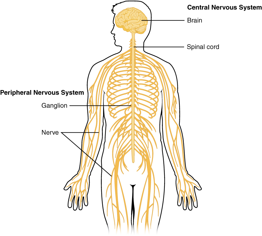
The picture you have in your mind of the nervous system probably includes the brain, the nervous tissue contained within the cranium, and the spinal cord, the extension of nervous tissue within the vertebral column. That suggests it is made of two organs—and you may not even think of the spinal cord as an organ—but the nervous system is a very complex structure. Within the brain, many different and separate regions are responsible for many different and separate functions. It is as if the nervous system is composed of many organs that all look similar and can only be differentiated using tools such as the microscope or electrophysiology .
Watch this video:
Media 19.1 The Nervous System, Part 1: Crash Course A&P #8 [Online video]. Copyright 2015 by CrashCourse.
Nervous System Medical Terms
Anatomy (Structures) of the Nervous System
The Central and Peripheral Nervous Systems
Nervous tissue, present in both the CNS and PNS, contains two basic types of cells: neurons and glial cells. Neurons are the primary type of cell that most anyone associates with the nervous system. They are responsible for the computation and communication that the nervous system provides. They are electrically active and release chemical signals to target cells. Glial cells, or glia, are known to
play a supporting role for nervous tissue. Ongoing research pursues an expanded role that glial cells might play in signalling, but neurons are still considered the basis of this function. Neurons are important, but without glial support they would not be able to perform their function. A glial cell is one of a variety of cells that provide a framework of tissue that supports the neurons and their activities. The neuron is the more functionally important of the two, in terms of the communicative function of the nervous system. To describe the functional divisions of the nervous system, it is important to understand the structure of a neuron.
Neurons are cells and therefore have a soma, or cell body, but they also have extensions of the cell; each extension is generally referred to as a process. There is one important process that every neuron has called an axon, which is the fiber that connects a neuron with its target. Another type of process that branches off from the soma is the dendrite. Dendrites are responsible for receiving most of the input from other neurons.
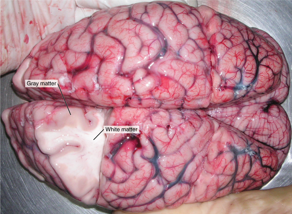
Looking at nervous tissue, there are regions that predominantly contain cell bodies and regions that are largely composed of just axons. These two regions within nervous system structures are often referred to as gray matter (the regions with many cell bodies and dendrites) or white matter (the regions with many axons). Figure 19.2 demonstrates the appearance of these regions in the brain and spinal cord. The colours ascribed to these regions are what would be seen in “fresh,” or unstained, nervous tissue. Gray matter is not necessarily gray. It can be pinkish because of blood content, or even slightly tan, depending on how long the tissue has been preserved. White matter is white because axons are insulated by a lipid-rich substance called myelin. Lipids can appear as white (“fatty”) material, much like the fat on a raw piece of chicken or beef. Actually, gray matter may have that colour ascribed to it because next to the white matter, it is just darker—hence, gray.
The distinction between gray matter and white matter is most often applied to central nervous tissue, which has large regions that can be seen with the unaided eye. When looking at peripheral structures, often a microscope is used and the tissue is stained with artificial colours. That is not to say that central nervous tissue cannot be stained and viewed under a microscope, but unstained tissue is most likely from the CNS—for example, a frontal section of the brain or cross section of the spinal cord.
The Adult Brain
The Cerebrum
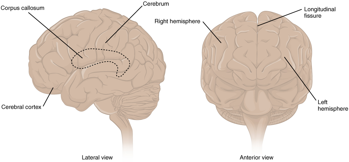
The iconic gray mantle of the human brain, which appears to make up most of the mass of the brain, is the cerebrum (see Figure 19.3). The wrinkled portion is the cerebral cortex, and the rest of the structure is beneath that outer covering. There is a large separation between the two sides of the cerebrum called the longitudinal fissure. It separates the cerebrum into two distinct halves, a right and left cerebral hemisphere. Deep within the cerebrum, the white matter of the corpus callosum provides the major pathway for communication between the two hemispheres of the cerebral cortex.
Many of the higher neurological functions, such as memory, emotion, and consciousness, are the result of cerebral function. The complexity of the cerebrum is different across vertebrate species. The cerebrum of the most primitive vertebrates is not much more than the connection for the sense of smell. In mammals, the cerebrum comprises the outer gray matter that is the cortex (from the Latin word meaning “bark of a tree”) and several deep nuclei that belong to three important functional groups. The basal nuclei are responsible for cognitive processing, the most important function being that associated with planning movements. The basal forebrain contains nuclei that are important in learning and memory. The limbic cortex is the region of the cerebral cortex that part of the limbic system, a collection of structures involved in emotion, memory, and behavior.
Cerebral Cortex
The cerebrum is covered by a continuous layer of gray matter that wraps around either side of the forebrain—the cerebral cortex. This thin, extensive region of wrinkled gray matter is responsible for the higher functions of the nervous system. A gyrus (plural = gyri) is the ridge of one of those wrinkles, and a sulcus (plural = sulci) is the groove between two gyri. The pattern of these folds of tissue indicates specific regions of the cerebral cortex.
The head is limited by the size of the birth canal, and the brain must fit inside the cranial cavity of the skull. Extensive folding in the cerebral cortex enables more gray matter to fit into this limited space. If the gray matter of the cortex were peeled off of the cerebrum and laid out flat, its surface area would be roughly equal to one square meter.
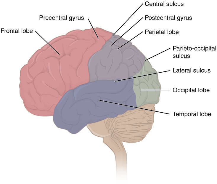
The folding of the cortex maximizes the amount of gray matter in the cranial cavity. During embryonic development, as the telencephalon expands within the skull, the brain goes through a regular course of growth that results in everyone’s brain having a similar pattern of folds. The surface of the brain can be mapped on the basis of the locations of large gyri and sulci. Using these landmarks, the cortex can be separated into four major regions, or lobes (see Figure 19.4). The lateral sulcus that separates the temporal lobe from the other regions is one such landmark. Superior to the lateral sulcus are the parietal lobe and frontal lobe, which are separated from each other by the central sulcus. The posterior region of the cortex is the occipital lobe, which has no obvious anatomical border between it and the parietal or temporal lobes on the lateral surface of the brain. From the medial surface, an obvious landmark separating the parietal and occipital lobes is called the parieto-occipital sulcus. The fact that there is no obvious anatomical border between these lobes is consistent with the functions of these regions being interrelated.
Concept Check
- Identify the two major divisions of the nervous system.
- Describe the cerebral cortex.
- What are the halves of the cerebrum know as?
The Diencephalon
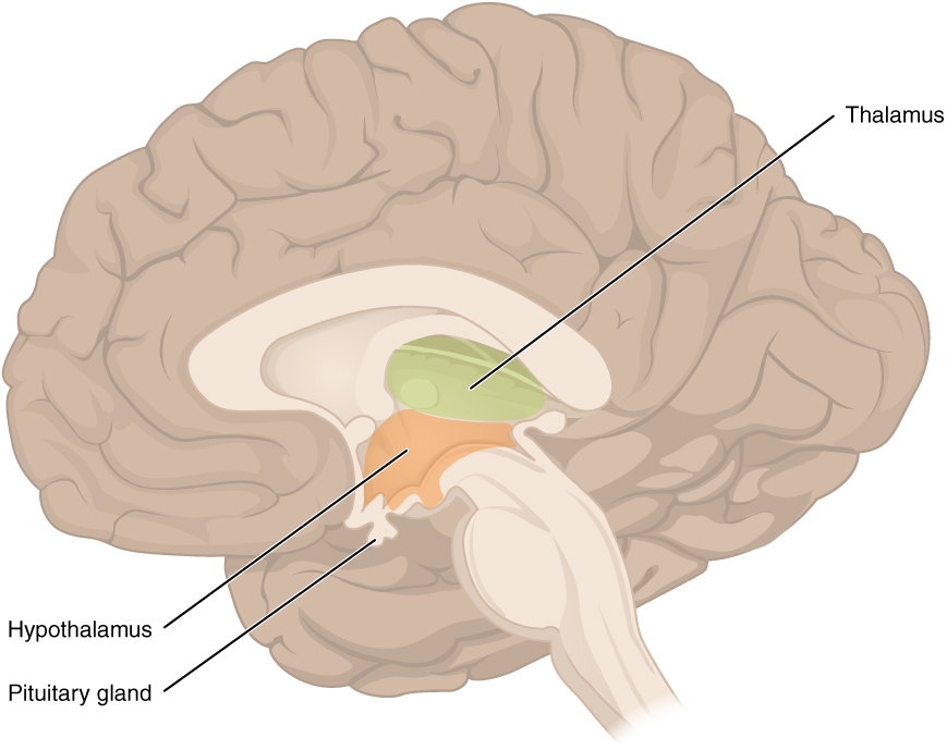
The diencephalon is deep beneath the cerebrum and constitutes the walls of the third ventricle. The diencephalon can be described as any region of the brain with “thalamus” in its name. The two major regions of the diencephalon are the thalamus itself and the hypothalamus (see Figure 19.5). There are other structures, such as the epithalamus, which contains the pineal gland, or the subthalamus, which includes the subthalamic nucleus that is part of the basal nuclei.
Thalamus
The thalamus is a collection of nuclei that relay information between the cerebral cortex and the periphery, spinal cord, or brain stem. All sensory information, except for the sense of smell, passes through the thalamus before processing by the cortex. For example, the portion of the thalamus that receives visual information will influence what visual stimuli are important, or what receives attention.
The cerebrum also sends information down to the thalamus, which usually communicates motor commands. This involves interactions with the cerebellum and other nuclei in the brain stem. The cerebrum interacts with the basal nuclei, which involves connections with the thalamus. The primary output of the basal nuclei is to the thalamus, which relays that output to the cerebral cortex. The cortex also sends information to the thalamus that will then influence the effects of the basal nuclei.
Hypothalamus
Inferior and slightly anterior to the thalamus is the hypothalamus, the other major region of the diencephalon. The hypothalamus is a collection of nuclei that are largely involved in regulating homeostasis. The hypothalamus is the executive region in charge of the autonomic nervous system and the endocrine system through its regulation of the anterior pituitary gland. Other parts of the hypothalamus are involved in memory and emotion as part of the limbic system.
Brain Stem
The midbrain and hindbrain (composed of the pons and the medulla) are collectively referred to as the brain stem (see Figure 19.6). The structure emerges from the ventral surface of the forebrain as a tapering cone that connects the brain to the spinal cord. Attached to the brain stem, but considered a separate region of the adult brain, is the cerebellum. The midbrain coordinates sensory representations of the visual, auditory, and somatosensory perceptual spaces. The pons is the main connection with the cerebellum. The pons and the medulla regulate several crucial functions, including the cardiovascular and respiratory systems and rates.
The cranial nerves connect through the brain stem and provide the brain with the sensory input and motor output associated with the head and neck, including most of the special senses. The major ascending and descending pathways between the spinal cord and brain, specifically the cerebrum, pass through the brain stem.
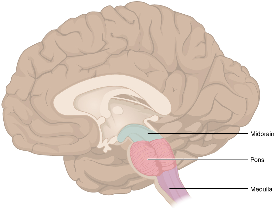
Midbrain
One of the original regions of the embryonic brain, the midbrain is a small region between the thalamus and pons. It is separated into the tectum and tegmentum, from the Latin words for roof and floor, respectively. The cerebral aqueduct passes through the center of the midbrain, such that these regions are the roof and floor of that canal.
Pons
The word pons comes from the Latin word for bridge. It is visible on the anterior surface of the brain stem as the thick bundle of white matter attached to the cerebellum. The pons is the main connection between the cerebellum and the brain stem. The bridge-like white matter is only the anterior surface of the pons; the gray matter beneath that is a continuation of the tegmentum from the midbrain. Gray matter in the tegmentum region of the pons contains neurons receiving descending input from the forebrain that is sent to the cerebellum.
Medulla
The medulla is the region known as the myelencephalon in the embryonic brain. The initial portion of the name, “myel,” refers to the significant white matter found in this region—especially on its exterior, which is continuous with the white matter of the spinal cord. The tegmentum of the midbrain and pons continues into the medulla because this gray matter is responsible for processing cranial nerve information. A diffuse region of gray matter throughout the brain stem, known as the reticular formation, is related to sleep and wakefulness, such as general brain activity and attention.
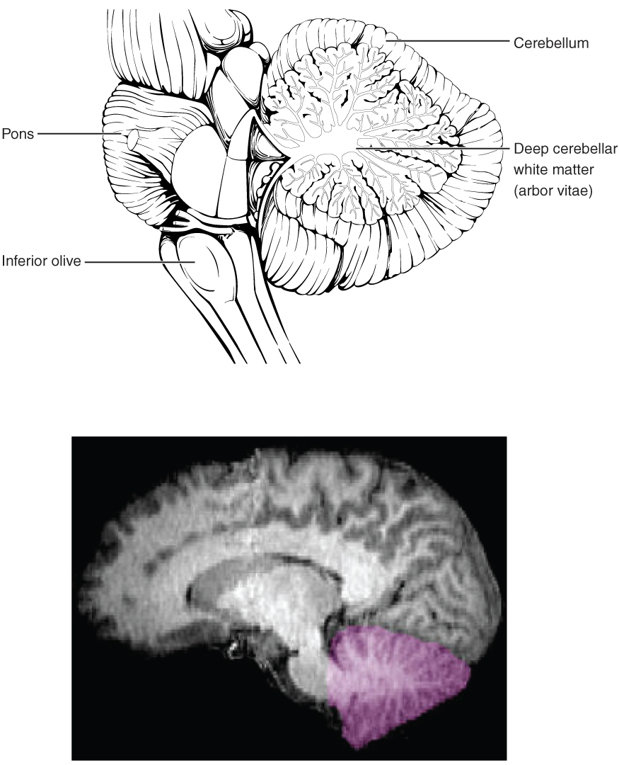
The Cerebellum
The cerebellum, as the name suggests, is the “little brain.” It is covered in gyri and sulci like the cerebrum, and looks like a miniature version of that part of the brain (see Figure 19.7). The cerebellum is largely responsible for comparing information from the cerebrum with sensory feedback from the periphery through the spinal cord. It accounts for approximately 10 percent of the mass of the brain.
Concept Check
- What is the primary processing purpose of the medulla?
- Identify the structure in the brain responsible for sensory feedback through the spinal cord. Suggest what may happen if this function failed.
The Spinal Cord
The description of the CNS is concentrated on the structures of the brain, but the spinal cord is another major organ of the system. Whereas the brain develops out of expansions of the neural tube into primary and then secondary vesicles, the spinal cord maintains the tube structure and is only specialized into certain regions. As the spinal cord continues to develop in the newborn, anatomical
features mark its surface. The anterior midline is marked by the anterior median fissure, and the posterior midline is marked by the posterior median sulcus. Axons enter the posterior side through the dorsal (posterior) nerve root, which marks the posterolateral sulcus on either side. The axons emerging from the anterior side do so through the ventral (anterior) nerve root. Note that it is common to see the terms dorsal (dorsal = “back”) and ventral (ventral = “belly”) used interchangeably with posterior and anterior, particularly in reference to nerves and the structures of the spinal cord. You should learn to be comfortable with both.
On the whole, the posterior regions are responsible for sensory functions and the anterior regions are associated with motor functions. This comes from the initial development of the spinal cord, which is divided into the basal plate and the alar plate. The basal plate is closest to the ventral midline of the neural tube, which will become the anterior face of the spinal cord and gives rise to motor neurons. The alar plate is on the dorsal side of the neural tube and gives rise to neurons that will receive sensory input from the periphery.
The length of the spinal cord is divided into regions that correspond to the regions of the vertebral column. The name of a spinal cord region corresponds to the level at which spinal nerves pass through the intervertebral foramina. Immediately adjacent to the brain stem is the following divisions of the spinal cord:
- cervical region
- thoracic region
- lumbar region
- sacral region
The spinal cord is not the full length of the vertebral column because the spinal cord does not grow significantly longer after the first or second year, but the skeleton continues to grow. The nerves that emerge from the spinal cord pass through the intervertebral formina at the respective levels. As the vertebral column grows, these nerves grow with it and result in a long bundle of nerves that resembles a horse’s tail and is named the cauda equina. The sacral spinal cord is at the level of the upper lumbar vertebral bones. The spinal nerves extend from their various levels to the proper level of the vertebral column.
Neurons
Neurons are the cells considered to be the basis of nervous tissue. They are responsible for the electrical signals that communicate information about sensations, and that produce movements in response to those stimuli, along with inducing thought processes within the brain. An important part of the function of neurons is in their structure, or shape. The three-dimensional shape of these cells makes the immense numbers of connections within the nervous system possible.
Parts of a Neuron
As you learned in the first section, the main part of a neuron is the cell body, which is also known as the soma (soma = “body”). The cell body contains the nucleus and most of the major organelles. But what makes neurons special is that they have many extensions of their cell membranes, which are generally referred to as processes. Neurons are usually described as having one, and only one, axon—a fiber that emerges from the cell body and projects to target cells. That single axon can branch repeatedly to communicate with many target cells. It is the axon that propagates the nerve impulse, which is communicated to one or more cells. The other processes of the neuron are dendrites, which receive information from other neurons at specialized areas of contact called synapses. The dendrites are usually highly branched processes, providing locations for other neurons to communicate with the cell body. Information flows through a neuron from the dendrites, across the cell body, and down the axon. This gives the neuron a polarity—meaning that information flows in this one direction. Figure 19.8 shows the relationship of these parts to one another.
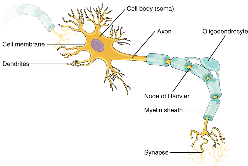
Where the axon emerges from the cell body, there is a special region referred to as the axon hillock. This is a tapering of the cell body toward the axon fiber. Within the axon hillock, the cytoplasm changes to a solution of limited components called axoplasm. Because the axon hillock represents the beginning of the axon, it is also referred to as the initial segment.
Many axons are wrapped by an insulating substance called myelin, which is actually made from glial cells. Myelin acts as insulation much like the plastic or rubber that is used to insulate electrical wires. A key difference between myelin and the insulation on a wire is that there are gaps in the myelin covering of an axon. Each gap is called a node of Ranvier and is important to the way that electrical signals travel down the axon. The length of the axon between each gap, which is wrapped in myelin, is referred to as an axon segment. At the end of the axon is the axon terminal, where there are usually several branches extending toward the target cell, each of which ends in an enlargement called a synaptic end bulb. These bulbs are what make the connection with the target cell at the synapse.
Glial Cells
Glial cells, or neuroglia or simply glia, are the other type of cell found in nervous tissue. They are considered to be supporting cells, and many functions are directed at helping neurons complete their function for communication. The name glia comes from the Greek word that means “glue,” and was coined by the German pathologist Rudolph Virchow, who wrote in 1856: “This connective substance, which is in the brain, the spinal cord, and the special sense nerves, is a kind of glue (neuroglia) in which the nervous elements are planted.” Today, research into nervous tissue has shown that there are many deeper roles that these cells play. And research may find much more about them in the future.
Myelin
Anatomy Labeling Activity
Return to the Table of Contents
Physiology (Function) of the Nervous System
The nervous system is involved in receiving information about the environment around us (sensation) and generating responses to that information (motor responses). The nervous system can be divided into regions that are responsible for sensation (sensory functions) and for the response (motor functions). But there is a third function that needs to be included. Sensory input needs to be integrated with other sensations, as well as with memories, emotional state, or learning (cognition). Some regions of the nervous system are termed integration or association areas. The process of integration combines sensory perceptions and higher cognitive functions such as memories, learning, and emotion to produce a response.
Sensation
The first major function of the nervous system is sensation—receiving information about the environment to gain input about what is happening outside the body (or, sometimes, within the body). The sensory functions of the nervous system register the presence of a change from homeostasis or a particular event in the environment, known as a stimulus. The senses we think of most are the “big five”: taste, smell, touch, sight, and hearing. The stimuli for taste and smell are both chemical substances (molecules, compounds, ions, etc.), touch is physical or mechanical stimuli that interact with the skin, sight is light stimuli, and hearing is the perception of sound, which is a physical stimulus similar to some aspects of touch. There are actually more senses than just those, but that list represents the major senses. Those five are all senses that receive stimuli from the outside world, and of which there is conscious perception. Additional sensory stimuli might be from the internal environment (inside the body), such as the stretch of an organ wall or the concentration of certain ions in the blood.
Response
The nervous system produces a response on the basis of the stimuli perceived by sensory structures. An obvious response would be the movement of muscles, such as withdrawing a hand from a hot stove, but there are broader uses of the term. The nervous system can cause the contraction of all three types of muscle tissue. For example, skeletal muscle contracts to move the skeleton, cardiac muscle is influenced as heart rate increases during exercise, and smooth muscle contracts as the digestive system moves food along the digestive tract. Responses also include the neural control of glands in the body as well, such as the production and secretion of sweat by the eccrine and merocrine sweat glands found in the skin to lower body temperature.
Responses can be divided into those that are voluntary or conscious (contraction of skeletal muscle) and those that are involuntary (contraction of smooth muscles, regulation of cardiac muscle, activation of glands). Voluntary responses are governed by the somatic nervous system and involuntary responses are governed by the autonomic nervous system, which are discussed in the next section.
Integration
Stimuli that are received by sensory structures are communicated to the nervous system where that information is processed. This is called integration. Stimuli are compared with, or integrated with, other stimuli, memories of previous stimuli, or the state of a person at a particular time. This leads to the specific response that will be generated. Seeing a baseball pitched to a batter will not automatically cause the batter to swing. The trajectory of the ball and its speed will need to be considered. Maybe the count is three balls and one strike, and the batter wants to let this pitch go by in the hope of getting a walk to first base. Or maybe the batter’s team is so far ahead, it would be fun to just swing away.
Controlling the Body
The nervous system can be divided into two parts mostly on the basis of a functional difference in responses. The somatic nervous system (SNS) is responsible for conscious perception and voluntary motor responses. Voluntary motor response means the contraction of skeletal muscle, but those contractions are not always voluntary in the sense that you have to want to perform them. Some somatic motor responses are reflexes, and often happen without a conscious decision to perform them. If your friend jumps out from behind a corner and yells “Boo!” you will be startled and you might scream or leap back. You didn’t decide to do that, and you may not have wanted to give your friend a reason to laugh at your expense, but it is a reflex involving skeletal muscle contractions. Other motor responses become automatic (in other words, unconscious) as a person learns motor skills (referred to as “habit learning” or “procedural memory”).
The autonomic nervous system (ANS) is responsible for involuntary control of the body, usually for the sake of homeostasis (regulation of the internal environment). Sensory input for autonomic functions can be from sensory structures tuned to external or internal environmental stimuli. The motor output extends to smooth and cardiac muscle as well as glandular tissue. The role of the autonomic system is to regulate the organ systems of the body, which usually means to control homeostasis. Sweat glands, for example, are controlled by the autonomic system. When you are hot, sweating helps cool your body down. That is a homeostatic mechanism. But when you are nervous, you might start sweating also. That is not homeostatic, it is the physiological response to an emotional state.
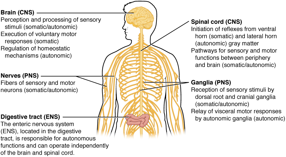
There is another division of the nervous system that describes functional responses. The enteric nervous system (ENS) is responsible for controlling the smooth muscle and glandular tissue in your digestive system. It is a large part of the PNS, and is not dependent on the CNS. It is sometimes valid, however, to consider the enteric system to be a part of the autonomic system because the neural structures that make up the enteric system are a component of the autonomic output that regulates digestion. There are some differences between the two, but for our purposes here there will be a good bit of overlap. See Figure 19.13 for examples of where these divisions of the nervous system can be found.
Functions of the Cerebral Cortex
The cerebrum is the seat of many of the higher mental functions, such as memory and learning, language, and conscious perception, which are the subjects of subtests of the mental status exam. The cerebral cortex is the thin layer of gray matter on the outside of the cerebrum. It is approximately a millimeter thick in most regions and highly folded to fit within the limited space of the cranial vault. These higher functions are distributed across various regions of the cortex, and specific locations can be said to be responsible for particular functions. There is a limited set of regions, for example, that are involved in language function, and they can be subdivided on the basis of the particular part of language function that each governs.
Cognitive Abilities
Assessment of cerebral functions is directed at cognitive abilities. The abilities assessed through the mental status exam can be separated into four groups: orientation and memory, language and speech, sensorium, and judgment and abstract reasoning.
Orientation and Memory
Orientation is the patient’s awareness of his or her immediate circumstances. It is awareness of time, not in terms of the clock, but of the date and what is occurring around the patient. It is awareness of place, such that a patient should know where he or she is and why. It is also awareness of who the patient is—recognizing personal identity and being able to relate that to the examiner. The initial tests of orientation are based on the questions, “Do you know what the date is?” or “Do you know where you are?” or “What is your name?” Further understanding of a patient’s awareness of orientation can come from questions that address remote memory, such as “Who is the President of the United States?”, or asking what happened on a specific date.
Memory is largely a function of the temporal lobe, along with structures beneath the cerebral cortex such as the hippocampus and the amygdala. The storage of memory requires these structures of the medial temporal lobe. A famous case of a man who had both medial temporal lobes removed to treat intractable epilepsy provided insight into the relationship between the structures of the brain and the function of memory.
The prefrontal cortex can also be tested for the ability to organize information. In one subtest of the mental status exam called set generation, the patient is asked to generate a list of words that all start with the same letter, but not to include proper nouns or names. The expectation is that a person can generate such a list of at least 10 words within 1 minute. Many people can likely do this much more quickly, but the standard separates the accepted normal from those with compromised prefrontal cortices.
Read this article to learn about a young man who texts his fiancée in a panic as he finds that he is having trouble remembering things. At the hospital, a neurologist administers the mental status exam, which is mostly normal except for the three-word recall test. The young man could not recall them even 30 seconds after hearing them and repeating them back to the doctor. An undiscovered mass in the mediastinum region was found to be Hodgkin’s lymphoma, a type of cancer that affects the immune system and likely caused antibodies to attack the nervous system. The patient eventually regained his ability to remember, though the events in the hospital were always elusive. Considering that the effects on memory were temporary, but resulted in the loss of the specific events of the hospital stay, what regions of the brain were likely to have been affected by the antibodies and what type of memory does that represent?
Language and Speech
Language is, arguably, a very human aspect of neurological function. There are certainly strides being made in understanding communication in other species, but much of what makes the human experience seemingly unique is its basis in language. Any understanding of our species is necessarily reflective, as suggested by the question “What am I?” And the fundamental answer to this question is suggested by the famous quote by René Descartes: “Cogito Ergo Sum” (translated from Latin as “I think, therefore I am”). Formulating an understanding of yourself is largely describing who you are to yourself. It is a confusing topic to delve into, but language is certainly at the core of what it means to be self-aware.
The neurological exam has two specific subtests that address language. One measures the ability of the patient to understand language by asking them to follow a set of instructions to perform an action, such as “touch your right finger to your left elbow and then to your right knee.” Another subtest assesses the fluency and coherency of language by having the patient generate descriptions of objects or scenes depicted in drawings, and by reciting sentences or explaining a written passage.
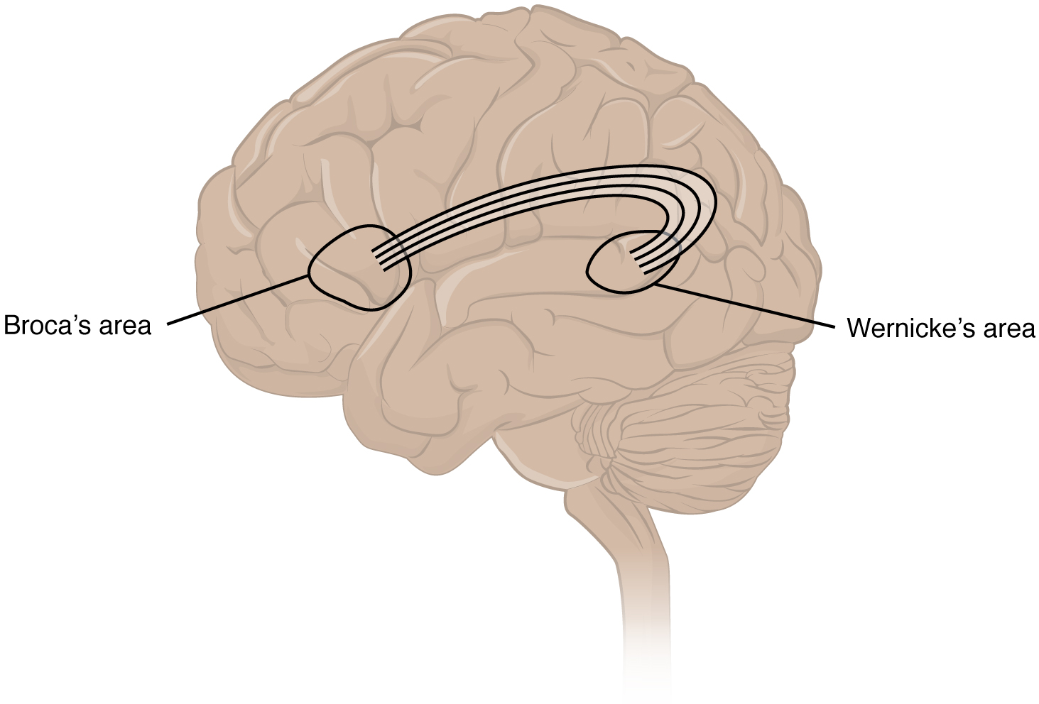
An important example of multimodal integrative areas is associated with language function (see Figure 19.14). Adjacent to the auditory association cortex, at the end of the lateral sulcus just anterior to the visual cortex, is Wernicke’s area. In the lateral aspect of the frontal lobe, just anterior to the region of the motor cortex associated with the head and neck, is Broca’s area. Both regions were originally described on the basis of losses of speech and language, which is called aphasia. The aphasia associated with Broca’s area is known as an expressive aphasia, which means that speech production is compromised. This type of aphasia is often described as non-fluency because the ability to say some words leads to broken or halting speech. Grammar can also appear to be lost. The aphasia associated with Wernicke’s area is known as a receptive aphasia, which is not a loss of speech production, but a loss of understanding of content. Patients, after recovering from acute forms of this aphasia, report not being able to understand what is said to them or what they are saying themselves, but they often cannot keep from talking.
The two regions are connected by white matter tracts that run between the posterior temporal lobe and the lateral aspect of the frontal lobe. Conduction aphasia associated with damage to this connection refers to the problem of connecting the understanding of language to the production of speech. This is a very rare condition, but is likely to present as an inability to faithfully repeat spoken language.
Sensorium
Those parts of the brain involved in the reception and interpretation of sensory stimuli are referred to collectively as the sensorium. The cerebral cortex has several regions that are necessary for sensory perception. Several of the subtests can reveal activity associated with these sensory modalities, such as being able to hear a question or see a picture. Two subtests assess specific functions of these cortical areas.
The first is praxis, a practical exercise in which the patient performs a task completely on the basis of verbal description without any demonstration from the examiner. The second subtest for sensory perception is gnosis, which involves two tasks. The first task, known as stereognosis, involves the naming of objects strictly on the basis of the somatosensory information that comes from manipulating them. The patient keeps their eyes closed and is given a common object, such as a coin, that they have to identify. The patient should be able to indicate the particular type of coin, such as a dime versus a penny, or a nickel versus a quarter, on the basis of the sensory cues involved. For example, the size, thickness, or weight of the coin may be an indication, or to differentiate the pairs of coins suggested here, the smooth or corrugated edge of the coin will correspond to the particular denomination. The second task, graphesthesia, is to recognize numbers or letters written on the palm of the hand with a dull pointer, such as a pen cap.
Judgment and Abstract Reasoning
The prefrontal cortex is responsible for the functions responsible for planning and making decisions. In the mental status exam, the subtest that assesses judgment and reasoning is directed at three aspects of frontal lobe function. First, the examiner asks questions about problem solving, such as “If you see a house on fire, what would you do?” The patient is also asked to interpret common proverbs, such as “Don’t look a gift horse in the mouth.” Additionally, pairs of words are compared for similarities, such as apple and orange, or lamp and cabinet.
Everyday Connections
Left Brain, Right Brain
Popular media often refer to right-brained and left-brained people, as if the brain were two independent halves that work differently for different people. This is a popular misinterpretation of an important neurological phenomenon. As an extreme measure to deal with a debilitating condition, the corpus callosum may be sectioned to overcome intractable epilepsy. When the connections between the two cerebral hemispheres are cut, interesting effects can be observed.
The reason for this is that the language functions of the cerebral cortex are localized to the left hemisphere in 95 percent of the population. Additionally, the left hemisphere is connected to the right side of the body through the corticospinal tract and the ascending tracts of the spinal cord. Motor commands from the precentral gyrus control the opposite side of the body, whereas sensory information processed by the postcentral gyrus is received from the opposite side of the body. For a verbal command to initiate movement of the right arm and hand, the left side of the brain needs to be connected by the corpus callosum. Language is processed in the left side of the brain and directly influences the left brain and right arm motor functions, but is sent to influence the right brain and left arm motor functions through the corpus callosum. Likewise, the left-handed sensory perception of what is in the left pocket travels across the corpus callosum from the right brain, so no verbal report on those contents would be possible if the hand happened to be in the pocket.
People who have had their corpus callosum cut can perform two independent tasks at the same time because the lines of communication between the right and left sides of his brain have been removed. Whereas a person with an intact corpus callosum cannot overcome the dominance of one hemisphere over the other, this patient can. If the left cerebral hemisphere is dominant in the majority of people, why would right-handedness be most common?
Common Nervous System Abbreviations
Return to the Table of Contents
Diseases, Disorders, and Conditions of the Skeletal System
Terms that Describe Types of Paralysis
| Term | Word Breakdown | Description |
|---|---|---|
| flaccid flAs-uhd |
flaccid flabby, pendulous, weak, drooping |
Lack of muscle tone; limp |
| hemiplegia hem-i-plEE-juh |
-plegia paralysis hemi- |
Paralysis that affects one side of the body. For example, the arm and leg on the same side of the body |
| hemiparesis hem-i-puhr-rEE-suhs |
-paresis weakness hemi- |
One sided weakness that affects one side of the body. For example, the arm and leg on the same side of the body |
| paraplegia pair-uh-plEE-jee-uh |
-plegia paralysis para- |
Paralysis that affects both legs and lower part of the body |
| paresis puhr-rEE-suhs |
-paresis weakness |
A partial paralysis wherein there is still some control of the muscles |
| quadriplegia kwah-druh-plEE-jee-uh |
-plegia paralysis quadri/o |
Paralysis that affects both arms, both legs and the central portion of the body |
| spastic spAs-tik |
-ic pertaining to spast/o |
Nervous System: Diseases, Conditions, & Disorders - Paralysis Term Word Breakdown Description flaccid flAs-uhd flaccid flabby, pendulous, weak, drooping Lack of muscle tone; limp hemiplegia hem-i-plEE-juh -plegia paralysis hemi- half Paralysis that affects one side of the body. For example, the arm and leg on the same side of the body hemiparesis hem-i-puhr-rEE-suhs -paresis weakness hemi- half One sided weakness that affects one side of the body. For example, the arm and leg on the same side of the body paraplegia pair-uh-plEE-jee-uh -plegia paralysis para- alongside, abnormal Paralysis that affects both legs and lower part of the body paresis puhr-rEE-suhs -paresis weakness A partial paralysis wherein there is still some control of the muscles quadriplegia kwah-druh-plEE-jee-uh -plegia paralysis quadri/o four Paralysis that affects both arms, both legs and sometimes from the neck down spastic spAs-tik -ic pertaining to spast/o |
To learn more about paralysis, please visit the Cleveland Clinic’s Paralysis information web page.
Return to the Table of Contents
Diseases, Conditions, & Disorders of the Brain
| Term | Word Breakdown | Description |
|---|---|---|
| amnesia am-nEE-zhuh |
-ia condition a- mnesis |
Loss of memories Read more |
| anencephaly an-en-sEf-uh-lee |
encephal/o brain an- |
When the brain is not fully formed. Read more |
| aphasia uh-fAY-zhuh |
-ia condition a- phas/o |
An impairment of language, affecting the production or comprehension of speech and the ability to read or write following a brain injury. Read more |
| cephalgia or cephalalgia | -algia pain cephal/o head |
Headache Infographics that address migraines |
| cerebrovascular accident (CVA) suhr-ree-broh-vAs-kyuh-luhr Ak-suh-duhnt |
-ar pertaining to cerebr/o vacul/o |
When a blood vessel that carries oxygen and nutrients to the brain is either blocked by a clot or bursts (or ruptures). When that happens, part of the brain cannot get the blood (and oxygen) it needs, so it and brain cells die. Read more |
| cerebral palsy | Cerebral palsy (CP) is a group of disorders that affect a person’s ability to move and maintain balance and posture. CP is the most common motor disability in childhood. Cerebral means having to do with the brain. Palsy means weakness or problems with using the muscles. CP is caused by abnormal brain development or damage to the developing brain that affects a person’s ability to control his or her muscles. Read more | |
| comatose kOH-muh-tohs |
-ose full of; pertaining to; sugar comat/o |
A deep state of unconsciousness. Information about rating the level of a coma. Glasgow Coma Scale |
| concussion kuhn-kUHsh-uhn |
ion- process concutere |
A type of traumatic brain injury—or TBI—caused by a bump, blow, or jolt to the head Video animation (30 seconds) Read more |
| dementia di-mEn-shu |
-ia condition de- ment/o |
A general term for loss of memory, language, problem-solving and other thinking abilities that are severe enough to interfere with daily life. Read more |
| dyslexia dis-lEk-see-uh |
-ia condition dys- lex/o |
A condition which causes challenges with reading and writing. People with dyslexia often experience challenges with other language skills such as spelling, writing, and pronouncing words. Read more |
| dysphasia dis-fAY-zhuh |
-ia condition phas/o dys- |
A condition that affects the ability to produce and understand spoken language. Difference between aphasia and dysphasia |
| encephalitis in-sef-uh-lIE-tuhs |
-itis
encephal/o |
Inflammation of the brain Read more |
| epilepsy Ep-uh-lep-see |
-lepsy seizure -epi |
A neurological disorder characterized by recurrent and unpredictable seizures. Read more |
| hematoma hee-muh-tOH-muh |
-oma tumor; mass; fluid collection hemat/o |
A collection a of blood outside of blood vessel An intracranial hematoma is bleed in the brain |
| hemorrhagic stroke | -ic pertaining to hem/o rrhage- |
A type of CVA (stroke) caused by the rupture of a blood vessel in the brain with bleeding into the surrounding tissue. Animation |
| hydrocephalus hie-droh-sEf-uh-luhs |
-us pertaining to hydr/o cephal/o |
An abnormal buildup of cerebrospinal fluid in the ventricles in the brain. This excess fluid causes the ventricles to widen, putting harmful pressure on the brain's tissues.Read more Video explanation (3 min 37 sec) |
| intracranial In-truh-krAY-nee-uhl |
-ar pertaining to crani/o intra- |
Within the skull |
| intraventricular in-truh-ven-trIk-yuh-luhr |
-ar pertaining to ventricul/o intra- |
Within the ventricles |
| ischemic stroke is-kEE-mik |
-emic pertaining to blood condition isk/o |
An interruption of blood flow to the brain that comes from a blockage, usually due to a blood clot or a stenosis (narrowing) of an artery that comes from atherosclerosis. Read more |
| meningitis men-uhn-jIE-tuhs |
-itis inflammation mening/o |
Inflammation of the meninges that surround and protect the brain and spinal cord. Bacterial meningitis is contagious and is a medical emergency. Read more |
| multiple sclerosis (MS) mUHl-tuh-puhl skluhr-rOH-suhs |
An autoimmune disease where the body attacks the myelin that covers the nerves. The results is signals don't travel efficiently from the brain to the receptor site, which affects muscle movement. Video explanation | |
| narcolepsy nAHR-kuh-lep-see |
-lepsy siezure narc/o |
People with narcolepsy are prone to falling asleep during everyday activity. Narcolepsy comes from a neurologic problem that affects sleep wake cycles. Read more |
| Parkinson's Disease pAHR-kuhn-suhnz di-zEEz |
A eponym is when a discovery is named after a patient or person. Parkinson's disease was named in 1817 by an English surgeon. | A progressive neurological disorder that primarily affects movement and is characterized by a range of motor and non-motor symptoms. Symptoms include, tremors, incoordination, slow movements, speech impairment, and sometimes cognitive decline. Read more |
| photophobia foh-tuh-fOH-bee-uh |
ia condition phot/o phob/o |
Abnormal light sensitivity |
| postictal | -al pertaining to post- ict/o |
Period of time after a seizure.
Neurologic symptoms may be evident as the person returns to a more normal state. |
| seizure sEE-zhuhr |
-ure (noun suffix, no meaning)
seize |
Episodes of electrical activity in the brain, which can alter or interrupt the transmission of signals between brain cells. Read more |
| syncope sIng-kuh-pee |
A loss of consciousness for a short period of time. Also called fainting or passing out. Comes from a sudden decrease of blood flow to the brain.Read more | |
| transient ischemic attack (TIA) | A temporary blockage of blood flow to the brain due to narrowing of artery that supplies blood to the brain. A TIA is a warning sign of a potential stroke. Animation |
Multiple sclerosis (MS) is one such disease. It is an example of an autoimmune disease. The antibodies produced by lymphocytes (a type of white blood cell) mark myelin as something that should not be in the body. This causes inflammation and the destruction of the myelin in the central nervous system. As the insulation around the axons is destroyed by the disease, scarring becomes obvious (Betts, et al., 2013).
Stroke
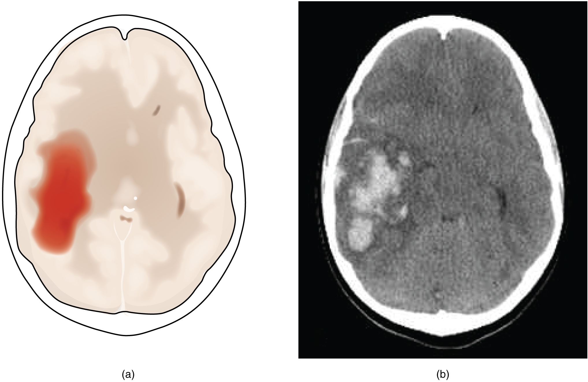
Damage to the nervous system can be limited to individual structures or can be distributed across broad areas of the brain and spinal cord. Localized, limited injury to the nervous system is most often the result of circulatory problems. The loss of blood flow to part of the brain is known as a stroke, or a cerebrovascular accident (CVA). There are two main types of stroke, depending on how the blood supply is compromised: ischemic and hemorrhagic. An ischemic stroke is the loss of blood flow to an area because vessels are blocked or narrowed. This is often caused by an embolus, which may be a blood clot or fat deposit. Ischemia may also be the result of thickening of the blood vessel wall, or a drop in blood volume in the brain known as hypovolemia. A hemorrhagic stroke is bleeding into the brain because of a damaged blood vessel. Accumulated blood fills a region of the cranial vault and presses against the tissue in the brain (see Figure 19.15) (Betts, et al., 2013).
Cerebral Palsy
Cerebral Palsy (CP) is caused by an interruption to the normal development of a person’s brain leading to weakness with muscles. Depending on the area of the brain that is affected, signs and symptoms will vary in the type and severity between individuals. Balance and coordination are often challenging due the inability to control muscles (Centers for Disease Control and Prevention, 2019, Ontario Federation for Cerebral Palsy, 2018). To learn more about cerebral palsy please visit the Centers for Disease Control and Prevention’s web page on cerebral palsy.
Traumatic Brain Injury (TBI)
According to the Minister of Health, approximately 20,000 people in Canada are hospitalized (each year) for traumatic brain injures. Brain injuries range from moderate to severe and include concussions. TBI can be caused by falls, automobile accidents, sports, assaults and strokes. Investment has been made to educate people on how to prevent TBIs with a focus on concussions from sports (Taylor, 2019).
Return to the Table of Contents
Diseases, Disorders & Conditions of the Spinal Cord
| Term | Word Breakdown | Description |
|---|---|---|
| meningocele | -cele bulge; hernia menig/o |
Part of the spinal cord to come through the spine like a sac that is pushed out. Nerve fluid is in the sac, and there is usually no nerve damage. Read more |
| meningomyelocele | -cele bulge; hernia menig/o myel/o |
When parts of the spinal cord and nerves come through the open part of the spine. It causes nerve damage and other disabilities. Read more |
| myelomeningocele | -cele bulge; hernia myel/o menig/o
|
Same as meningomyelocele |
| neural tube defect nUR-uhl tOOb EE-fekt |
Congenital problems that occur when the neural tube does not close properly. Leads to birth defects such as spina bifida and anencephaly. Read more | |
| spinal cord injury (SCI) spIE-nl kORd In-juhr-ree |
Damage to the spinal cord that causes motor and sensory issues from the point of injury downward. Read more |
Return to the Table of Contents
Diseases, Disorders & Conditions of the Nerves
| Term | Word Breakdown | Description |
|---|---|---|
| carpal tunnel syndrome kAHR-puhl tUHn-l sIn-drohm |
Disorder that occurs when the median nerve, which runs from your forearm into the palm of the hand, becomes pressed or squeezed at the wrist. Carpal Tunnel Syndrome causes pain, muscle weakness and numbness in the hand. Read more |
|
| causalgia kaw-zAl-juh |
-algia pain caus/o |
Burning sensation of a peripheral nerve |
| demyelination dee-mie-uh-luh-nAY-shuhn |
-tion process myelin/o de- |
Demyelination is a deterioration of the myelin sheath that covers the nerves. As a result messages sent along axons are slower which causes the axon to deteriorate. Demyelination interrupts signals from the brain and can cause affect motor and sensory systems. |
| neuralgia nu-rAl-juh |
-algia pain neur/o |
Nerve pain |
| neuritis nu-rIE-tuhs |
-itis inflammation neur/o |
Inflammation of more than one peripheral nerve that can cause pain, tingling, numbness and muscle weakness, and atrophy |
| neuroma nu-rOH-muh |
-oma tumor; mass; fluid collection neur/o |
A benign growth of nerve tissue. that causes pain. A Morton's neuroma causes pain between the toes. Read more |
| neuropathy nu-rAH-puh-thee |
-pathy disease neur/o |
Damage to the peripheral nerves that interrupts the signals from the central nervous system, The result can be moto or sensory disturbances. Read more |
| paresthesia par-uhs-thEE-zhuh |
-ia condition para- esthes/o |
Burning or itching feeling along a nerve. Also know as "pins and needles." Read more |
| polyneuritis pah-lee-nu-rIE-tuhs |
-itis inflammation neur/o nerve |
Inflammation of more than one peripheral nerve that can cause pain, tingling, numbness and muscle weakness, and atrophy |
| radiculopathy or radiculitis | -pathy disease radicul/o |
Pain, tingling, numbness and muscle weakness, and atrophy that comes from a compressed nerve. Also called a pinched nerve |
| sciatica | A condition characterized by pain, numbness, and/or weakness that radiates along the path of the sciatic nerve, which runs from the lower back through the hips and down each leg. |
Procedures Related to the Nervous System
| Term | Word Breakdown | Description |
|---|---|---|
| angiography an-jee-AH-gruh-fee |
-graphy process of recording angi/o |
Cerebral angiography is a process of creating a contrast x-ray of the blood vessels and blood flow in your brain. |
| myelography | -graphy process of recording spinal cord OR bone marrow |
Process of creating a contrast x-ray of the spinal cord, nerve roots and spinal lining (meninges) |
| electroencephalography(EEG) i-lek-troin-sef-uh-lAH-gruh-fee |
-graphy process of recording electr/o encephal/o |
The use small electric sensors to measure brain activity and nerve function |
| polysomnography | -graphy process of recording poly- somn/o |
The process of recording brain waves, oxygen levels, and heart rate during sleep. Also known as a sleep study. |
| positron emission tomography (PET) scan pAH-zuh-trahn ee-mIsh-uhn toh-mAH-gruh-fee |
A PET scan of the brain shows both structures and functions within the brain. |
Electroencephalogram (EEG)
With electrodes applied to your scalp, an EEG measures electrical activity in the brain. It’s used to help diagnose conditions of the brain, including inflammation, tumors, and injuries, as well as seizures and psychiatric disorders.
Return to the Table of Contents
Surgical Procedures
| Term | Word Breakdown | Description |
|---|---|---|
| craniotomy kray-nee-AH-tuh-mee |
-otomy incision; to cut into crani/o |
A surgery to cut an opening in the skull. Read more |
| diskectomy | -ectomy cut out; surgical removal disk |
The surgical removal of the damaged portion of a herniated disk in the spine |
| laminectomy | -ectomy cut out; surgical removal lamin/o |
Surgical removal of part or all of the vertebral bone (lamina). Laminectomy may be used to remove bone spurs or a herniated (slipped) disk in your spine. The procedure can take pressure off your spinal nerves or spinal cord. Read more |
| lumbar puncture (LP) lUHm-buhr pUHngk-chuhr |
Puncture of the lumbar vertebrae. Conducted 1) to extract a small amount of cerebral spinal fluid with a special needle. Used to diagnose medical conditions. or 2) to administer medication. Read more | |
| spondylodesis | -desis surgical binding spndyl/o |
Also known as spinal fusion. Procedure to fuse unstable vertebrae. |
Lumbar Puncture
Return to the Table of Contents
Drug Categories
| Term | Word Breakdown | Description |
|---|---|---|
| anticonvulsant an-tee-kuhn-vUHl-suhnt |
-anti
against |
Prevents seizures (another word for antiepileptic) |
| antiepileptic | -ic pertaining to anti- epi leps/o |
Prevents seizures (another word for anticonvulsant) |
| narcotic nahr-kAH-tik |
-ic
pertaining to narc/o numbness; stupor; sleep |
Also known as an opioid. Used to control severe pain by dulling the senses and relieve pain. Prescription opioids are generally safe when taken for a short time and as directed by a doctor, but because they produce euphoria in addition to pain relief, they can be misused and have addiction potential. Read more |
| Term | Word Breakdown | Description |
|---|---|---|
| neurologist nu-rAH-luh-jist |
-ist specialist neur/o nerve |
physician skilled in the diagnosis and treatment of disease of the nervous system |
| occupational therapist/occupational therapy assistant | Occupational therapists and occupational therapy assistants focus on the things you want and need to do in your daily life. Occupational therapy intervention uses everyday life activities (occupations) to promote health, well-being, and your ability to participate in the important activities in your life. This includes any meaningful activity that a person wants to accomplish, including taking care of yourself and your family, working, volunteering, going to school, among many others. American Occupational Therapy Association) |
Primary Specialist – Neurologist
Neurologists are medical doctors who complete an additional 5years of specialized training in the prevention, diagnosis, and treatment of disorders and conditions related to the brain, spinal cord, nerves and muscles (Canadian Medical Association, 2018).. For more details please follow the link to the Canadian Medical Association’s page on Neurology profile (PDF file).
Occupational Therapy Assistant Program at Tulsa Community College
Tulsa Community Community College has an Occupational Therapy Assistant program that prepares graduates to sit for the national certification exam and practice occupational therapy under the supervision of an occupational therapist. Read more about the program
Read more about the profession
References
Bergen, T. (2018). Tensilon test. Healthline. https://www.healthline.com/health/tensilon-test
Canadian Cancer Society. (2020). Lumbar puncture. https://www.cancer.ca/en/cancer-information/diagnosis-and-treatment/tests-and-procedures/lumbar-puncture/?region=on
Centers for Disease Control and Prevention. (2018). Mental health. CDC. https://www.cdc.gov/mentalhealth/learn/index.htm
Centers for Disease Control and Prevention. (2019). TBI: Get the facts. CDC. https://www.cdc.gov/conjunctivitis/about/causes.html
Centers for Disease Control and Prevention. (2020). Cerebral palsy (CP). CDC. https://www.cdc.gov/conjunctivitis/about/causes.html
Cherney, K. & De Pietro, M. (2019). Neurologist. Healthline. https://www.healthline.com/find-care/articles/neurologists/neurologist
[CrashCourse]. (2015, Feburary 23). The nervous system, part 1: Crash course A&P #8 [Video]. YouTube. https://www.youtube.com/watch?v=qPix_X-9t7E
Merriam-Webster. (n.d.). Neurologist. In Merriam-Webster.com dictionary. https://www.merriam-webster.com/dictionary/neurologist
Moores, D., & Cirino, E. (2018). Electromyography (EMG). Healthline. https://www.healthline.com/health/electromyography
Ontario Federation for Cerebral Palsy. (2018). About cerebral palsy. https://www.ofcp.ca/about-cerebral-palsy
Taylor, G. (2019, June 3). Brain injury awareness month – June 2019. Public Health Agency of Canada. https://www.canada.ca/en/public-health/news/2019/05/brain-injury-awareness-month–june-2019.html
Image Descriptions
Figure 19.1 image description: This diagram shows a silhouette of a human highlighting the nervous system. The central nervous system is composed of the brain and spinal cord. The brain is a large mass of ridged and striated tissue within the head. The spinal cord extends down from the brain and travels through the torso, ending in the pelvis. Pairs of enlarged nervous tissue, labeled ganglia, flank the spinal cord as it travels through the rib area. The ganglia are part of the peripheral nervous system, along with the many thread-like nerves that radiate from the spinal cord and ganglia through the arms, abdomen and legs. [Return to Figure 19.1].
Figure 19.2 image description: This photo shows an enlarged view of the dorsal side of a human brain. The right side of the occipital lobe has been shaved to reveal the white and gray matter beneath the surface blood vessels. The white matter branches though the shaved section like the limbs of a tree. The gray matter branches and curves on outside of the white matter, creating a buffer between the outer edges of the occipital lobe and the internal white matter. [Return to Figure 19.2].
Figure 19.3 image description: This figure shows the lateral view on the left panel and anterior view on the right panel of the brain. The major parts including the cerebrum are labeled. Lateral view labels (clockwise from top) read: cerebrum, cerebral cortex, corpus callosum (located on the interior of the brain). Anterior view labels indicate the right and left hemispheres, and the longitudinal fissure between them. [Return to Figure 19.3].
Figure 19.4 image description: This figure shows the lateral view of the brain and the major lobes are labeled. From the front of the brain (left) labels read: frontal lobe, precentral gyrus, central sulcus, postcentral gyrus, parietal lobe, pateral sulcus, occipital lobe, temporal lobe. [Return to Figure 19.4].
Figure 19.5 image description: This figure shows the location of the thalamus, hypothalamus and pituitary gland in the brain. Each part is labelled respectively. The thalamus is located in the midsection of the brain. The hypothalamus is located below the thalamus, and the pituitary gland below that. [Return to Figure 19.5].
Figure 19.6 image description: This figure shows the location of the midbrain, pons and the medulla in the brain that make up the brain stem. The midbrain is located at the top, the pons is located beneath that, and the medulla is the lowest most point of the brain stem. [Return to Figure 19.6].
Figure 19.7 image description: This figure shows the location of the cerebellum in the brain which is located on the posterior surface of the brain stem. Labels read (top, left): pons, inferior olive, (top, right) cerebellum, deep cerebellar white matter (arbor vitae). In the top panel, a lateral view labels the location of the cerebellum and the deep cerebellar white matter. In the bottom panel, a photograph of a brain, with the cerebellum in pink is shown. [Return to Figure 19.7].
Figure 19.8 image description: This illustration shows the anatomy of a neuron. The neuron has a very irregular cell body (soma) containing a purple nucleus. There are six projections protruding from the top, bottom and left side of the cell body. Each of the projections branches many times, forming small, tree-shaped structures protruding from the cell body. The right side of the cell body tapers into a long cord called the axon. The axon is insulated by segments of myelin sheath, which resemble a semitransparent toilet paper roll wound around the axon. The myelin sheath is not continuous, but is separated into equally spaced segments. The bare axon segments between the sheath segments are called nodes of Ranvier. An oligodendrocyte is reaching its two arm like projections onto two myelin sheath segments. The axon branches many times at its end, where it connects to the dendrites of another neuron. Each connection between an axon branch and a dendrite is called a synapse. The cell membrane completely surrounds the cell body, dendrites, and its axon. The axon of another nerve is seen in the upper left of the diagram connecting with the dendrites of the central neuron. [Return to Figure 19.8].
Figure 19.9 image description: Three illustrations show some of the possible shapes that neurons can take. In the unipolar neuron, the dendrite enters from the left and merges with the axon into a common pathway, which is connected to the cell body. The axon leaves the cell body through the common pathway, the branches off to the right, in the opposite direction as the dendrite. Therefore, this neuron is T shaped. In the bipolar neuron, the dendrite enters into the left side of the cell body while the axon emerges from the opposite (right) side. In a multipolar neuron, multiple dendrites enter into the cell body. The only part of the cell body that does not have dendrites is the part that elongates into the axon. [Return to Figure 19.9].
Figure 19.10 image description: This diagram contains three black and white drawings of more specialized nerve cells. Part A shows a pyramidal cell of the cerebral cortex, which has two, long, nerve tracts attached to the top and bottom of the cell body. However, the cell body also has many shorter dendrites projecting out a short distance from the cell body. Part B shows a Purkinje cell of the cerebellar cortex. This cell has a single, long, nerve tract entering the bottom of the cell body. Two large nerve tracts leave the top of the cell body but immediately branch many times to form a large web of nerve fibers. Therefore, the purkinje cell somewhat resembles a shrub or coral in shape. Part C shows the olfactory cells in the olfactory epithelium and olfactory bulbs. It contains several cell groups linked together. At the bottom, there is a row of olfactory epithelial cells that are tightly packed, side-by-side, somewhat resembling the slats on a fence. There are six neurons embedded in this epithelium. Each neuron connects to the epithelium through branching nerve fibers projecting from the bottom of their cell bodies. A single nerve fiber projects from the top of each neuron and synapses with nerve fibers from the neurons above. These upper neurons are cross shaped, with one nerve fiber projecting from the bottom, top, right and left sides. The upper cells synapse with the epithelial nerve cells using the nerve tract projecting from the bottom of their cell body. The nerve tract projecting from the top continues the pathway, making a ninety degree turn to the right and continuing to the right border of the image. [Return to Figure 19.10].
Figure 19.11 image description: This diagram shows several types of nervous system cells associated with two multipolar neurons. Astrocytes are star shaped-cells with many dendrite like projections but no axon. They are connected with the multipolar neurons and other cells in the diagram through their dendrite like projections. Ependymal cells have a teardrop shaped cell body and a long tail that branches several times before connecting with astrocytes and the multipolar neuron. Microglial cells are small cells with rectangular bodies and many dendrite like projections stemming from their shorter sides. The projections are so extensive that they give the microglial cell a fuzzy appearance. The oligodendrocytes have circular cell bodies with four dendrite like projections. Each projection is connected to a segment of myelin sheath on the axons of the multipolar neurons. The oligodendrocytes are the same color as the myelin sheath segment and are adding layers to the sheath using their projections. [Return to Figure 19.11].
Figure 19.12 image description: This diagram shows a collection of PNS glial cells. The largest cell is a unipolar peripheral ganglionic neuron which has a common nerve tract projecting from the bottom of its cell body. The common nerve tract then splits into the axon, going off to the left, and the dendrite, going off to the right. The cell body of the neuron is covered with several satellite cells that are irregular, flattened, and take on the appearance of fried eggs. Schwann cells wrap around each myelin sheath segment on the axon, with their nucleus creating a small bump on each segment. [Return to Figure 19.12].
Figure 19.13 image description: A silhouette of a human with only the brain, spinal cord, PNS ganglia, nerves and a section of the digestive tract visible. The brain, which is part of the CNS, is the area of perception and processing of sensory stimuli (somatic/autonomic), the execution of voluntary motor responses (somatic), and the regulation of homeostatic mechanisms (autonomic). The spinal cord, which is part of the CNS, is the area where reflexes are initiated. The gray matter of the ventral horn initiates somatic reflexes while the gray matter of the lateral horn initiates autonomic reflexes. The spinal cord is also the somatic and autonomic pathway for sensory and motor functions between the PNS and the brain. The nerves, which are part of the PNS, are the fibers of sensory and motor neurons, which can be either somatic or autonomic. The ganglia, which are part of the PNS, are the areas for the reception of somatic and autonomic sensory stimuli. These are received by the dorsal root ganglia and cranial ganglia. The autonomic ganglia are also the relay for visceral motor responses. The digestive tract is part of the enteric nervous system, the ENS, which is located in the digestive tract and is responsible for autonomous function. The ENS can operate independent of the brain and spinal cord. [Return to Figure 19.13].
Figure 19.15 image description: The left panel of this figure shows an image of the brain with a region in red. This red region indicates a hemorrhage associated with a stroke. The right panel shows a hemorrhage as it might appear on a CT scan. [Return to Figure 19.15].
Return to the Table of Contents
Unless otherwise indicated, this chapter contains material adapted from Anatomy and Physiology (on OpenStax), by Betts, et al. and is used under a a CC BY 4.0 international license. Download and access this book for free at https://openstax.org/books/anatomy-and-physiology/pages/1-introduction.
instrument to view small particles by enlarging the particles
The study of electrical properties of cells and tissues
includes the brain and spinal cord
all nervous tissue that is outside of the brain and spinal cord
region of the adult brain that is responsible for higher neurological functions such as memory, emotion, and consciousness
a collection of nucleic nerve tissue - has function in both the autonomic and endocrine systems - regulates homeostasis
