14 Female Reproductive System
Learning Objectives
- Identify the anatomy of the female reproductive system
- Describe the main functions of the female reproductive system
- Spell the medical terms of the female reproductive system and use correct abbreviations
- Identify the medical specialties associated with the female reproductive system
Chapter Fourteen: Table of Contents
What Can Go Wrong? – Diseases, Disorders, and Conditions of the Female Reproductive System
How Do We Fix it or Make it Better?
References, Attributions, and Image Descriptions
Introduction to the Female Reproductive System
| Term | Word Breakdown | Description |
|---|---|---|
| breast brEst |
The female breast is mostly made up of a collection of fat cells called adipose tissue. Milk ducts in the breast carry milk to the nipple. National Breast Cancer Foundation | |
| cervix sUHR-viks |
cervic/o neck; cervix (neck of uterus) |
Lower part of the uterus that connects to the vagina. The cervix has a small opening that expands during childbirth. It also allows menstrual blood to leave a woman's body. Medline Plus |
| fallopian tubes fuh-lOH-pee-uhn tOObz |
The ovaries are connected to the uterus by the uterine tubes (fallopian tubes). The egg travels through the tube to the uterus. Fertilization occurs in the fallopian tubes. Medline Plus | |
| ovaries OH-vuhr-reez |
Female reproductive organs responsible for producing eggs and hormones. | |
| ovulation ah-vyuh-lAY-shuhn |
Process where that includes the development of a mature egg and release from the ovary. | |
| peritoneum pair-uh-tuh-nEE-uhm |
-um structure; tissue; thing peritone/o |
A thin, transparent membrane that lines the abdominal cavity and covers most of the organs within it. In the female reproductive system, the peritoneum plays an important role in supporting and protecting the reproductive organs, including the uterus, ovaries, fallopian tubes, and vagina. |
| uterus yOO-tuhr-ruhs |
-us structure thing uter/o |
Hollow organ that houses and nourishes a developing fetus. Medline Plus |
| vagina vuh-jIE-nuh |
vagin/o vagina |
Muscular canal connecting the uterus to the external genitalia. |
The female reproductive system produces gametes and reproductive hormones. In addition, the female reproductive system supports the developing fetus and delivers it to the outside world. The female reproductive system is located primarily inside the pelvic cavity. The female gonads are called ovaries and the gamete they produce is called an oocyte.
Return to the Table of Contents
Female Reproductive System Word Parts
Click on prefixes, combining forms, and suffixes to reveal a list of word parts to memorize for the female reproductive system.
Return to the Table of Contents
Watch this video:
Media 10.1. Reproductive System, Part 1 – Female Reproductive System: Crash Course A&P #40 [Online video]. Copyright 2015 by CrashCourse.
Female Reproductive System Medical Terms
Return to the Table of Contents
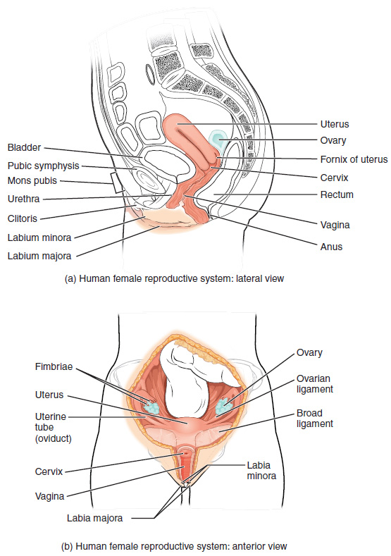
Anatomy (Structures) of the Female Reproductive System
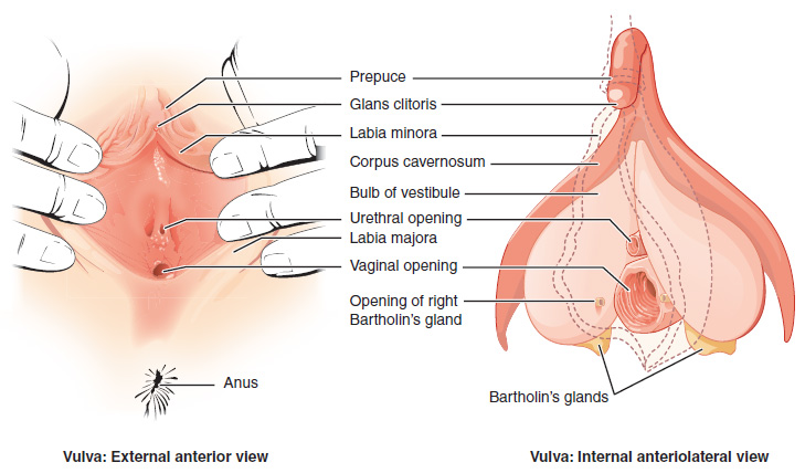
External Female Genitals
The external female reproductive structures are referred to collectively as the vulva and they include:
- The mons pubis is a pad of fat that is located at the anterior, over the pubic bone. After puberty, it becomes covered in pubic hair.
- The labia majora (labia = “lips”; majora = “larger”) are folds of hair-covered skin that begin just posterior to the mons pubis.
- The labia minora (labia = “lips”; minora = “smaller”) is thinner and more pigmented and extends medially to the labia majora.
- Although they naturally vary in shape and size from woman to woman, the labia minora serve to protect the female urethra and the entrance to the female reproductive tract.
- The superior, anterior portions of the labia minora come together to encircle the clitoris (or glans clitoris), an organ that originates from the same cells as the glans penis and has abundant nerves that make it important in sexual sensation and orgasm. The hymen is a thin membrane that sometimes partially covers the entrance to the vagina.
- The vaginal opening is located between the opening of the urethra and the anus. It is flanked by outlets to the Bartholin’s glands.
Internal Female Reproductive Organs
Vagina
The vagina is a muscular canal (approximately 10 cm long) that is the entrance to the reproductive tract. It also serves as the exit from the uterus during menses and childbirth. The outer walls of the anterior and posterior vagina are columns with ridges. The superior fornix meets the uterine cervix. The cervix is the opening to the uterus.
The walls of the vagina are lined with:
- An outer, fibrous adventitia
- A middle layer of smooth muscle
- An inner mucous membrane with transverse folds called rugae.
Together, the middle and inner layers allow the expansion of the vagina to accommodate intercourse and childbirth. The thin, perforated hymen can partially surround the opening to the vaginal orifice. The Bartholin’s glands and the lesser vestibular glands (located near the clitoris) secrete mucus, which keeps the vestibular area moist.
The vagina has a normal population of microorganisms that help to protect against infection. There is both pathogenic bacteria, and yeast in the vagina. In a healthy woman, the most predominant type of vaginal bacteria is from the genus Lactobacillus, which secretes lactic acid. the lactic acid protects the vagina by maintaining an acidic pH (below 4.5).
Lactic acid, in combination with other vaginal secretions, makes the vagina a self-cleansing organ. However, douching can disrupt the normal balance of healthy microorganisms, and increase a woman’s risk for infections and irritation. It is recommend that women do not douche and that they allow the vagina to maintain its normal healthy population of protective microbial flora.
Ovaries
The ovaries are the female gonads. There are two, one at each entrance to the fallopian tube. They are each about 2 to 3 cm in length, about the size of an almond. The ovaries are located within the pelvic cavity. The ovary itself is attached to the uterus via the ovarian ligament. The ovarian stroma forms the bulk of the adult ovary. Oocytes develop within the outer layer of this stroma, each surrounded by supporting cells. This grouping of an oocyte and its supporting cells is called a follicle.
The Fallopian Tubes
The fallopian tubes are the conduit of the oocyte from the ovary to the uterus. Each of the two fallopian tubes is close to, but not directly connected to, the ovary.
- The isthmus is the narrow medial end of each uterine tube that is connected to the uterus.
- The wide distal infundibulum flares out with slender, finger-like projections called fimbriae.
- The middle region of the tube, called the ampulla, is where fertilization often occurs.
The fallopian tubes have three layers:
- An outer serosa
- A middle smooth muscle layer
- An inner mucosal layer
- In addition to its mucus-secreting cells, the inner mucosa contains ciliated cells that beat in the direction of the uterus, producing a current that will be critical to moving the oocyte .
The Uterus and Cervix
The uterus is the muscular organ that nourishes and supports the growing embryo. Its average size is approximately 5 cm wide by 7 cm long and it has three sections.
- The portion of the uterus superior to the opening of the uterine tubes is called the fundus.
- The middle section of the uterus is called the body of uterus (or corpus).
- The cervix is the narrow inferior portion of the uterus that projects into the vagina.
- The cervix produces mucus secretions that become thin and stringy under the influence of high systemic plasma estrogen concentrations, and these secretions can facilitate sperm movement through the reproductive tract.
The wall of the uterus is made up of three layers:
- Perimetrium: the most superficial layer and serous membrane.
- Myometrium: a thick layer of smooth muscle responsible for uterine contractions.
- Endometrium: the innermost layer containing a connective tissue lining covered by epithelial tissue that lines the lumen. It provides the site of implantation for a fertilized egg, and sheds during menstruation if no egg is fertilized.
Concept Check
- Write or draw out the components of the pathway that an oocyte takes from beginning to end.
- Why do you think the fallopian tubes are not connected to the ovaries?
Return to the Table of Contents
Physiology (Function) of the Female Reproductive System-Ovulation
Following ovulation, the Fallopian tube receives the oocyte. Oocytes lack flagella, and therefore cannot move on their own.
- High concentrations of estrogen that occur around the time of ovulation induce contractions of the smooth muscle along the length of the Fallopian tube.
- These contractions occur every 4 to 8 seconds, causing the oocyte to flow towards the uterus, through the coordinated beating of the cilia that line the outside and lumen of the length of the Fallopian tube which pulls the oocyte into the interior of the tube.
- Once inside, the muscular contractions and beating cilia move the oocyte slowly toward the uterus.
- When fertilization does occur, sperm typically meet the egg while it is still moving through the ampulla.
Watch this video:
Media 10.2. Ovulation. From Betts, et al., 2013. Licensed under CC BY 4.0.
The Menstrual Cycle
The three phases of the menstrual cycle are:
- The menses phase of the menstrual cycle is the phase during which reproductive hormone levels are low, the woman menstruates, and the lining is shed. The menses phase lasts between 2 – 7 days with an average of 5 days.
- The proliferative phase is when menstrual flow ceases and the endometrium begins to proliferate . During this phase reproductive hormones are working in homeostasis to trigger ovulation on approximately day 14 of a typical 28-day menstrual cycle. Ovulation marks the end of the proliferative phase.
- The secretory phase the endometrial lining prepares for implantation of a fertilized egg. If no pregnancy occurs within approximately 10- 12 days the endometrium will grow thinner and shed starting the first day of the next cycle.
Anatomy Labeling Activity
Female Reproductive System Terms not Easily Broken into Word Parts
Female Reproductive System Medical Abbreviations
Return to the Table of Contents
Diseases, Disorders and Conditions of the Female Reproductive System
Diseases, Disorders and Conditions of the Breast
| Term | Word Breakdown | Description |
|---|---|---|
| breast cancer brEst kAn-suhr |
Breast cancer is a cancer that starts in breast tissue. It happens when cells in the breast change and grow out of control. The cells usually form a tumor.
Sometimes the cancer does not spread any further. This is called "in situ." If the cancer spreads outside the breast, the cancer is called "invasive." It may just spread to nearby tissues and lymph nodes. Or the cancer may metastasize (spread to other parts of the body) through the lymph system or the blood. |
|
| fibrocystic breasts fie-bruh-sIs-tik brEsts |
-ic pertaining to fibr/o cyst/o |
Fibrocystic breasts are painful, lumpy breasts. Formerly called fibrocystic breast disease, this common condition is, in fact, not a disease. Many women experience these normal breast changes, usually around their period. Medline Plus |
| galactorrhea guh-lak-tuhr-rEE-uh |
-rrha flow or discharge galact/o |
Breast milk production that is not related to childbirth. Medline Plus |
Return to the Table of Contents
Diseases, Disorders and Conditions of the Ovaries and Fallopian Tubes
| Term | Word Breakdown | Description |
|---|---|---|
| anovulation | -ation process; condition an- ovul/o |
When a woman doesn’t ovulate (release a fully developed egg) during a menstrual cycle, it’s called anovulation. CDC Infertility |
| polycystic ovary syndrome (PCOS) pah-lee-sIs-tik OH-vuhr-ree sIn-drohm |
-ic pertaining to poly- cyst/o |
A condition in which a woman has increased levels of male hormones (androgens). Many problems occur as a result of this increase of hormones, including:
Menstrual irregularities In many women with PCOS, mature eggs are not released. Instead, they stay in the ovaries with a small amount of fluid (cyst) around them. The affected ovary may be slightly enlarged. There can be many of these. However, not all women with the condition will have ovaries with this appearance. Medline Plus |
| salpingitis sal-puhn-jIE-tuhs Also known as pelvic inflammatory disease (PID) |
-itis inflammation salping/o |
PID refers to an infection of the uterus, fallopian tubes, or ovaries. |
Return to the Table of Contents
Diseases, Disorders and Conditions of the Uterus and Cervix
| Term | Word Breakdown | Description |
|---|---|---|
| endometriosis en-doh-mee-tree-OH-suhs |
-osis condition endo- metri/o |
A condition in which the tissue that normally grows inside the uterus grows outside of it |
| leiomyoma | -oma tumor; mass; fluid collection lei/o my/o |
Benign tumor of the smooth muscle of the uterus. Also known as fibroids |
| leiomyosarcoma | -oma tumor; mass; fluid collection lei/o my/o sarc/o |
A malignant tumor of the smooth muscle of the uterus |
| myometritis | -itis inflammation my/o metr/o |
Inflammation of the muscular layer of the uterus |
| pyometra | -ia condition -a noun ending py/o pus metr/o |
A condition characterized by the presence of pus in the uterus |
Return to the Table of Contents
Diseases, Disorders and Conditions of the Vagina and Peritoneum
| Term | Word Breakdown | Description |
|---|---|---|
| candidiasis kan-duh-dIE-uh-suhs |
-iasis condition, formation of |
An infection caused by a yeast (a type of fungus) called Candida. Symptoms include: vaginal itching or soreness; pain during sexual intercourse; Pain or discomfort when urinating; and abnormal vaginal discharge. CDC - Vaginal Candidiasis |
| leukorrhea | -rrhea flow or discharge leuk/o |
Vaginal discharge, typically whitish or yellowish in color. Can be caused from an estrogen imbalance, a sexually transmitted disease, or a yeast infection. Healthline |
| rectocele | -cele bulge; hernia retr/o |
Condition in which the rectum protrudes into the back wall of the vagina. Can cause pelvic pain. difficulties with defecation, constipation, and dyspareunia. National Institute of Health |
Return to the Table of Contents
Menstrual Disorders
| Term | Word Breakdown | Description |
|---|---|---|
| amenorrhea ay-men-uhr-rEE-uh |
-rrhea flow or discharge a- men/o |
The absence of menstrual periods in a woman of reproductive age |
| dysmenorrhea dis-men-uhr-rEE-uh |
-rrhea
dys- men/o |
Painful menstruation |
| menopause mEn-uh-pawz |
-pause
men/o |
The natural cessation of menstrual periods and fertility in women, typically occurring around the age of 50 |
| menorrhagia men-uhr-rAY-juh |
-rrahgia bursting forth (of blood) men/o |
A condition characterized by abnormally heavy or prolonged menstrual bleeding |
| oligomenorrhea uh-li-gu-meh-nr-ee-uh |
-rrhea flow or discharge oligo- men/o |
Abnormally light or infrequent menstrual periods. |
Return to the Table of Contents
Diagnostic Procedures related to the Female Reproductive System
Female Reproductive System: Diagnostic Procedures
| Term | Word Breakdown | Description |
|---|---|---|
| colposcopy kahl-pAH-skuh-pee |
-scopy process of visually examining colp/o |
Procedure that uses a colposcope to examine the cervix, vagina, and vulva. Medline Plus - Colposcopy |
| hysterosalpingography his-tuhr-roh-sal-ping-gAH-gruh-fee |
-graphy process of recording hyster/o salping/o |
Radiographic examination that looks at the shape of the uterus and checks whether the fallopian tubes are open. Radiological Society of North America, Inc. (RSNA). |
| mammography ma-mAH-gruh-fee |
-graphy process of recording mamm/o |
X-ray imaging to screen and detect breast abnormalities Medline Plus - Mammography |
Return to the Table of Contents
Surgical Procedures related to the Female Reproductive System
| Term | Word Breakdown | Description |
|---|---|---|
| colporrhaphy | -rrhaphy suture colp/o |
Surgical repair of the vaginal wall |
| cryosurgery krie-oh-sUHRj-ree |
-surgery
cry/o |
A technique that uses extreme cold to destroy abnormal or diseased tissue. |
| hysterectomy his-tuhr-rEk-tuh-mee |
-ectomy cut out; surgical removal hyster/o |
The surgical removal of the uterus. |
| oophorectomy oh-uh-fuhr-rEk-tuh-mee |
-ectomy cut out; surgical removal oophor/o |
Surgical removal of one or both ovaries |
| lumpectomy luhm-pEk-tuh-mee |
-ectomy cut out; surgical removal |
The surgical removal of a breast tumor and a small margin of surrounding healthy tissue |
| mammaplasty | -plasty surgical repair mamm/o |
Surgical reconstruction of a breast to restore its shape and appearance? |
| mastectomy ma-stEk-tuh-mee |
-ectomy cut out; surgical removal mast/o |
Surgical removal of one or both breasts. |
| salpingectomy | -ectomy cut out; surgical removal salping/o |
Surgical removal of one or both fallopian tubes. |
Hysterectomy
A hysterectomy is done to stage or treat female reproductive cancers, treat precancerous conditions of the cervix and some non-cancerous conditions that have not responded to other forms of treatment. There are three types of hysterectomy:
- A total hysterectomy removes both the uterus and the cervix.
- A subtotal hysterectomy removes the uterus only.
- A radical hysterectomy removes uterus, cervix, part of the vagina, and ligaments.
Sometimes the ovaries and fallopian tubes are removed at the same time that a hysterectomy is done. A bilateral salpino-oophorectomy (BSO) removes both ovaries and fallopian tubes. A unilateral salpingo-oophorectomy removes one ovary and one Fallopian tube (Canadian Cancer Society, 2020). To learn more about hysterectomy please follow visit the Canadian Cancer Society’s page on hysterectomies.
Return to the Table of Contents
Female Reproductive System Medical Abbreviations
Return to the Table of Contents
Medical Terms in Context
Return to the Table of Contents
Medical Specialties
| Term | Word Breakdown | Description |
|---|---|---|
| gynecologist gie-nuh-kAH-luh-jist |
-logist specialist gynec/o |
A physician who specializes in the female reproductive system. |
Gynecology
A gynecologist is a specialist in the area of gynecology focusing on the diagnosis, treatment, management and prevention of diseases and disorders of the female reproductive system. Obstetrics is a specialty that provides care through pregnancy, labour, and puerperium. Further subspecialties in women’s health include contraception, reproductive endocrinology, infertility, adolescent gynecology, endoscopy and gynecological oncology (Canadian Medical Association, 2018). To learn more about obstetrics or gynecology please follow visit the Canadian Medical Association’s Obstetrics/Gynecology Profile page (PDF file).
Return to the Table of Contents
References
Canadian Medical Association. (2018, August). Obstetrics/gynecology profile. Canadian Medical Association Specialty Proflies. https://www.cma.ca/sites/default/files/2019-01/obgyn-e.pdf
[CrashCourse]. (2015, October 2015). Reproductive system, part 1 – female reproductive system: Crash course A&P #40 [Video]. YouTube. https://www.youtube.com/watch?v=RFDatCchpus
Return to the Table of Contents
Image Descriptions
Figure 10.1 image description: This figure shows the structure and the different organs in the female reproductive system. The top panel shows the lateral view with labels (clockwise from top): utuerus, ovary, formix of uterus, cervix, rectum, vagina, anus, labium majora, labium minora, clitoris, urethra, mons pubis, pybic symphysis, bladder; and the bottom panel shows the anterior view with labels (clockwise from top): ovary, ovarian ligament, broad ligament, labia minora, labia majora, vagina, cervix, uterine tube, usterus, fimbriae. [Return to Figure 10.1].
Figure 10.2 image description: This figure shows the parts of the vulva. The right panel shows the external anterior view and the left panel shows the internal anteriolateral view. The major parts are labeled (from top): prepuce, glans clitoris, labia minora, corpus cavernosum, bulb of vestibule, urethral opening, labia majora, vaginal opening, opening of right Bartholin’s gland, Bartholin’s glands, anus. [Return to Figure 10.2].
Return to the Table of Contents
Unless otherwise indicated, this chapter contains material adapted from Anatomy and Physiology (on OpenStax), by Betts, et al. and is used under a a CC BY 4.0 international license. Download and access this book for free at https://openstax.org/books/anatomy-and-physiology/pages/1-introduction.
haploid reproductive cells that contribute genetic material to form an offspring
superior portion of the vagina
Also known as greater vestibular glands they are responsible to secrete mucus to keep the vestibular area moist
washing the vagina with fluid
female gamete
pertaining to above
pertaining to below
reproduce rapidly
biological process that results in stable equilibrium
Surgical removal of the uterus
pertaining to both sides
pertaining to one side
Specialist in the study and treatment of the female reproductive system
The study of the female reproductive system
Time directly after childbirth
The study of endocrine glands and hormones
Process of viewing internally
