10 Muscular System
Learning Objectives
- Identify the anatomy of the muscular system
- Describe the main functions of the muscular system
- Spell the medical terms of the muscular system and use correct abbreviations
- Explore common diseases, disorders, and procedures related to the muscular system
- Identify the medical specialties associated with the muscular system
Chapter Ten: Table of Contents
What is it?
What Can Go Wrong? – Diseases, Disorders, and Conditions of the Muscular System
- Symptoms
- Specific Diseases, Disorders and Conditions of the Muscle
- Diseases, Disorders and Conditions of the Fascia, or Tendon
- Movement Disorders
How Do We Fix it or Make it Better?
References, Attributions, and Image Descriptions
Introduction to the Muscular System
| Term | Word Breakdown | Description |
|---|---|---|
| fascia fAY-shee-uh |
A sheet of connective tissue covering or binding together body structures (such as muscles) Read more | |
| ligament lIg-uh-muhnt |
ligament/o ligament |
Ligaments bind bone to bone and create stability of joints |
| muscle | muscl/o muscle |
A bundle of long slender cells (muscle fibers) that have the power to contract and hence to produce movement. Muscles are responsible for locomotion and play an important part in performing vital body functions. They also protect the contents of the abdomen against injury and help support the body. |
| tendon | tendin/o tendon |
Tendons connect muscles to bones. Made of fibrous tissue and collagen, tendons are tough but not very stretchy. (Cleveland Clinic) |
| agonist | A muscle whose contraction moves a part of the body directly. | |
| antagonist | An antagonist muscle is a muscle that produces the opposite action of an agonist The antagonist provides stability. | |
| cardiac muscle | These muscles line the heart walls. They help your heart pump blood that travels through your cardiovascular system. You don’t control cardiac muscles. Your heart tells them when to contract. (Cleveland Clinic) | |
| skeletal muscle | Also known as striated muscles. Skeletal muscles are responsible for voluntary muscle movement. | |
| smooth muscle | Also known as visceral muscles. Smooth muscle is mainly associated with the walls of internal organs. Smooth muscles are responsible for involuntary muscle movement. |
Muscular System Word Parts
Click on prefixes, combining forms, and suffixes to reveal a list of word parts to memorize for the Muscular System.
When most people think of muscles, they think of the muscles that are visible just under the skin, particularly of the limbs. These are skeletal muscles, so-named because most of them move the skeleton. But there are two additional types of muscles: the smooth muscle and the cardiac muscle. The body has over 600 muscles which contribute significantly to the body’s weight.
Return to the Table of Contents
Watch this video:
Media 17.1 Muscles, Part 2 – Organismal Level: Crash Course A&P #22 [Online video]. Copyright 2015 by CrashCourse.
Muscular System Medical Terms
Anatomy (Structures) of the Muscular System
Muscle is one of the four primary tissue types of the body, and it is made up of specialized cells called fibers. The body contains three types of muscle tissue: skeletal muscle, cardiac muscle, and smooth muscle (see Figure 17.1). All three muscle tissues have some properties in common; they all exhibit a quality called excitability as their plasma membranes can change their electrical states (from polarized to depolarized) and send an electrical wave called an action potential along the entire length of the membrane. Fascia is fibrous connective tissue that encloses muscles.
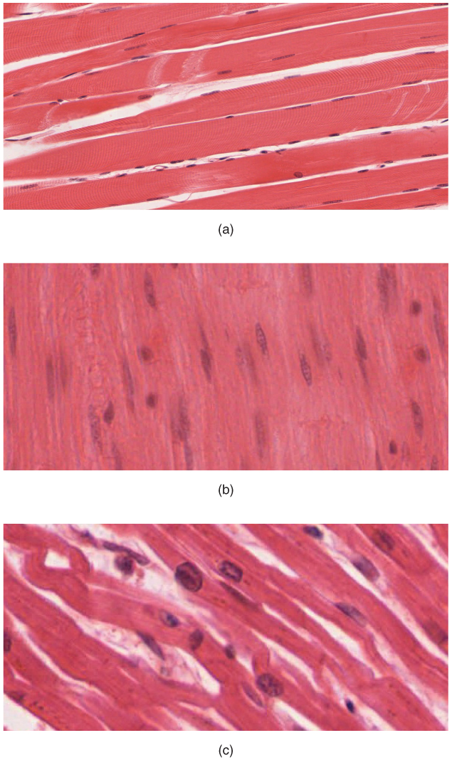
Three Types of Muscle Tissues
- Skeletal – closely associated with the skeletal system. Also known as striated muscles and are responsible for voluntary muscle movement – such as swallowing, etc.
- Smooth – mainly associated with the walls of the internal organs. Also known as visceral muscles and are responsible for involuntary muscle movement – such as breathing, etc.
- Cardiac – heart muscle or myocardium. Its appearance is similar to a skeletal muscle and is responsible for the pumping of blood. It gives the heart beat.
Skeletal Muscle
Skeletal muscles act not only to produce movement but also to stop movement, such as resisting gravity to maintain posture. Small, constant adjustments of the skeletal muscles are needed to hold a body upright or balanced in any position. Muscles also prevent excess movement of the bones and joints, maintaining skeletal stability and preventing skeletal structure damage or deformation.
Skeletal muscles are located throughout the body at the openings of internal tracts to control the movement of various substances. These muscles allow functions, such as swallowing, urination, and defecation, to be under voluntary control. Skeletal muscles also protect internal organs (particularly abdominal and pelvic organs) by acting as an external barrier or shield to external trauma and by supporting the weight of the organs.
Skeletal muscles contribute to the maintenance of homeostasis in the body by generating heat. This heat is very noticeable during exercise, when sustained muscle movement causes body temperature to rise, and in cases of extreme cold, when shivering produces random skeletal muscle contractions to generate heat.
Smooth Muscle
Smooth muscle, so named because the cells do not have striations, is present in the walls of hollow organs like the urinary bladder, uterus, stomach, intestines, and in the walls of passageways, such as the arteries and veins of the circulatory system, and the tracts of the respiratory, urinary, and reproductive systems. Smooth muscle is also present in the eyes, where it functions to change the size of the iris and alter the shape of the lens; and in the skin where it causes hair to stand erect in response to cold temperature or fear.
Cardiac Muscle
Cardiac muscle tissue is only found in the heart. Highly coordinated contractions of cardiac muscle pump blood into the vessels of the circulatory system. Similar to skeletal muscle, cardiac muscle is striated and organized into sarcomeres, possessing the same banding organization as skeletal muscle (see Figure 17.1). Cardiac muscle fibers cells also are extensively branched and are connected to one another at their ends by intercalated discs. An intercalated disc allows the cardiac muscle cells to contract in a wave-like pattern so that the heart can work as a pump.
Concept Check
- Compare and contrast the 3 types of muscles tissues.
- Where in the body do you find each of the muscle types?
Return to the Table of Contents
Physiology (Function) of the Muscular System
The main function of the muscular system is to assist with movement. Muscles work as antagonistic pairs. As one muscle contracts, the other muscle relaxes. This contraction pulls on
the bones and assists with movement. Contraction is the shortening of the muscle fibers while relaxation lengthens the fibers. This sequence of relaxation and contraction is influenced by the nervous system.
Muscles also work to keep the posture of the body. This is done through muscle contraction where the trunk is kept straight either when sitting or standing.
Naming of Muscles
There are many nomenclatures for naming muscles. Some of these include:
- divisions – biceps, triceps, quadriceps
- size – maximus (largest), minimus (smallest)
- shape – deltoid (triangular), trapezious (trapezoid)
- action – flexor (to flex), adductor (towards midline of body)
| EXAMPLE | WORD | LATIN ROOT 1 | LATIN ROOT 2 | MEANING | TRANSLATION |
| abductor digiti minimi | abductor | ab = away from | duct = to move | a muscle that moves away from | A muscle that moves the little finger or toe away |
| digiti | digitus = digit | n/a | refers to a finger or toe | ||
| minimi | minimus = mini, tiny | n/a | little | ||
| adductor digiti minimi | adductor | ad = to, toward | duct = to move | a muscle that moves towards | A muscle that moves the little finger or toe toward |
| digiti | digitus = digit | n/a | refers to a finger or toe | ||
| minimi | minimus = mini, tiny | n/a | little |
Diseases, Disorders, and Conditions of the Muscular System
Muscular System – Symptoms
| Term | Word Breakdown | Description |
|---|---|---|
| atrophy At-ruh-fee |
-trophy growth a- |
Decrease in size or wasting away of a body part or tissue |
| contracture kuhn-trAk-chuhr |
-ure result contract/o |
A condition in which a muscle or group of muscles remains in a state of permanent contraction Read more |
| hypertrophy hie-pUHR-truh-fee |
-trophy growth hyper- |
Excessive Growth. Hypertrophy is a muscle is the enlargement of muscles |
| strain strAYn |
An overstretching or overexertion of a muscle or tendon. | |
| myalgia mie-Al-juh |
-algia pain my/o |
Muscle pain |
| polymyalgia pah-lee-mie-Al-juh |
-algia pain poly my/o |
Muscle pain that affects several muscle groups |
| myopathy mie-AH-puh-thee |
-pathy disease my/o |
Diseases that attack muscle fibers, making your muscles weak. |
| myositis mie-uh-sIE-tuhs |
-sitis
myos/o |
Inflammation or infection of a muscle |
| polymyositis pah-lee-mie-uh-sIE-tuhs |
poly -many, excessive my/o |
Inflammation or infection of multiple muscle groups |
Sprain and Strain
A sprain is an injury to a joint whereby a ligament is stretched or torn.
A strain is an injury to a muscle whereby a tendon is stretched or torn.
Return to the Table of Contents
Conditions, Disorders & Diseases of the Muscle
| Term | Word Breakdown | Description |
|---|---|---|
| avulsion uh-vUHl-shuhn |
An avulsion fracture is where a small piece of bone attached to a tendon or ligament gets pulled away from the main part of the bone. Read more | |
| fibromyalgia fie-broh-mie-Al-juh |
-algia pain fibr/o my/o |
Fibromyalgia causes bodywide pain in the fibrous tissues of the muscles and extreme tiredness. It can be confused with arthritis because it may cause pain in joints, muscles and soft tissues Read more |
| myasthenia gravis My-as-theen-ee-a grav-u |
asthenia weakness my/o astheni/o |
Grave or serious muscle weakness. A rare neuromuscular, autoimmune disorder. Causes extreme fatigue and profound muscle weakness. Impulses from the brain travel down nerves but are blocked by antibodies before impulses reach muscle. The body is essentially attacking itself and stopping muscle function. Can impact a person’s ability to see, swallow, smile, walk, breathe, or engage in normal, everyday activity. Read more |
| muscular dystrophy mUHs-kyuh-luhr dIs-truh-fee |
-trophy growth. dys- |
The muscular dystrophies are a group of diseases that cause weakness and degeneration of the skeletal muscles. Read more |
| rhabdomyolysis rab-doh-mie-AH-luh-suhs |
-lysis break down, destruction, dissolving rhabd/o my/o |
Rhabdomyolysis is a rare muscle injury where your muscles break down. This is a life-threatening condition that can happen after an injury or excessive exercise without rest. Read more |
| rhabdomyoma rAb-doh-mie-OH-muh |
-oma tumor; mass; fluid collection rhabd/o my/o |
A a benign tumor of striated muscle. |
| rhabdomyosarcoma | -oma tumor; mass; fluid collection rhabd/o my/o sarc/o |
Rhabdomyosarcoma (RMS) is a rare type of cancer that forms in soft tissue — specifically skeletal muscle tissue or sometimes hollow organs such as the bladder or uterus. RMS can occur at any age, but it most often affects children Read more |
| sarcopenia | -penia deficiency; lack of sarc/o |
The age-related progressive loss of muscle mass and strength. |
| torticollis tor-tuh-kAH-luhs |
collis neck tortus |
Torticollis occurs when there is a contracture on one side of the neck that causes the head to turn and rotate to one side. Common in infants.Read more |
Duchenne Muscular Dystrophy
Duchenne Muscular Dystrophy (DMD) is caused by the inability of the body to make dystrophin (a muscle protein). This causes the muscles to become weak as the person ages. This disease primarily effects boys and signs and symptoms typically present before the age of five. Signs and symptoms may include frequent falls and trouble keeping up with peers. Since all muscles are affected, the person will eventually require a wheelchair and assistance with breathing (Muscular Dystrophy Canada, 2020). To learn more please visit Muscular Dystrophy Canada’s neuromuscular disorders web page.
Return to the Table of Contents
Conditions Disorders & Disease of the Fascia, or Tendon
| Term | Word Breakdown | Description |
|---|---|---|
| fasciitis fash-ee-IE-tuhs |
-itis inflammation or infection fasci/o |
Inflammation of the fascia. Plantar fasciitis involves inflammation of a thick band of tissue that runs across the bottom of each foot and connects the heel bone to the toes, known as the plantar fascia. |
| tendinitis ten-duh-nIE-tuhs |
-itis inflammation or infection tendin/o tendon |
Tendonitis is when a tendon is inflamed. It can happen to any tendon in the body. When a tendon is inflamed, it can cause swelling, pain, and discomfort. Read more |
| tenosynovitis tEn-oh-sin-uh-vIE-tuhs |
-itis inflammation or infection ten/o synov/o |
Inflammation of the lining of the tendon sheath around a tendon. |
Return to the Table of Contents
Movement Disorders
| Term | Word Breakdown | Description |
|---|---|---|
| ataxia uh-tAk-see-uh |
-ia condition a- tax/o |
Ataxia is a nervous system disorder that leads to poor muscle coordination during movement. Ataxia mimic those of being drunk, such as slurred speech, stumbling, falling, and incoordination. Read more |
| bradykinesia | -ia condition brady- kines/i |
Condition of slow movement. |
| dyskinesia dis-kuh-nEE-zhuh |
-ia condition dys- kines/i |
Abnormal motions due to difficulty controlling the voluntary muscles. |
| hyperkinesis hie-puhr-kuh-nEE-suhs |
-ia condition hyper- kines/i |
Condition of excessive movement. |
| myoclonus mie-AH-kluh-nuhs |
my/o muscle clonus |
Myoclonus refers to sudden, brief involuntary twitching or jerking of a muscle or group of muscles. The twitching cannot be stopped or controlled by the person experiencing it. Myoclonus is not a disease itself, rather it describes a clinical sign. Read more |
Return to the Table of Contents
Muscle Tests
| Term | Word Breakdown | Description |
|---|---|---|
| electromyogram i-lek-troh-mIE-uh-gram |
-gram record or picture electr/o my/o |
A record or display produced by electromyography. The electromyogram is the record that is produced during electromyography |
| electromyography i-lek-troh-mie-AH-gruh-fee |
-graphy process of recording electr/o my/o |
Electromyography (EMG) is a diagnostic test that evaluates the health and function of your skeletal muscles and the nerves that control them. |
| magnetic resonance imaging (MRI) mag-nEt-ik rEz-uh-nuhns Im-ij-ing |
Test that uses radio frequency waves and a magnetic field to produce clear images that aid in the diagnosis of a wide range of conditions (London Health Sciences Centre, 2020). |
Return to the Table of Contents
Electromyography (EMG) is a procedure that assesses the function of nerve cells that control muscles. Electrodes, either attached to the skin or inserted into the muscle, allow for the recording of electrical impulses. EMG can indicate functional problems with the peripheral nerves, muscles, or with the signals between the nerves and the muscles. This is just one test in a series of tests that assist in the diagnosis of neuromuscular disorders (Mayo Clinic Staff, 2019; Body Restoration, 2020). To learn more, please visit the Mayo Clinic’s Electromyography web page.
Magnetic Resonance Imaging (MRI) is a test that uses radio frequency waves and a magnetic field to produce clear images that aid in the diagnosis of a wide range of conditions (London Health Sciences Centre, 2020). Leung (2017) notes that there has been increased clinical use in using MRI for the treatment and monitoring of muscular disorders due to the high-quality MRI images that distinguish skeletal muscles from fat (para. 4).
Return to the Table of Contents
Muscular System: Surgical Procedures
| Term | Word Breakdown | Description |
|---|---|---|
| fasciectomy (fash-ee-EK-tuh-mee) |
-ectomy cut out; removal fasci/o |
A surgical procedure to remove fascia |
| fasciotomy (fash-ee-ec-toe-mee |
-otomy Incision fasci/o |
A procedure in which the fascia is cut to relieve pressure in the muscle compartment |
| myorrhaphy migh-or-rha-fee |
-rrhaphy suture my/o |
Suture of a muscle |
| tenomyoplasty ten-OH-my-OH-past-ee |
-plasty surgical repair ten/o my/o |
Surgical procedure involving the repair of both muscle and tendon |
| tenorrhaphy ten-OR-uh-fee |
-rrhaphy suture ten/o |
Suture of a tendon |
Muscular System: Drug Categories
| Term | Word Breakdown | Description |
|---|---|---|
| neuromuscular blocker drugs | Drugs that interrupt transmission of nerve impulses at the skeletal neuromuscular junction, Used in surgery to inhibit breathing which is then temporarily provided through mechanical ventilation. Read more | |
| relaxant | Drugs that relieve muscle spasm and stiffness Read more |
Return to the Table of Contents
Medical Specialties Related to Muscular System
| Term | Word Breakdown | Description |
|---|---|---|
| physiatrist fiz-ee-At-rist |
-ist specialist iatr/o physi/o |
Physiatry is the medical specialty that emphasizes the prevention, diagnosis, treatment and rehabilitation of people disabled by disease, disorder or injury. |
| physical therapist or physical therapy assistant (PT/PTA) | -ist specialist therap/o -al physic/o |
Physical therapists optimize quality of life by maintaining, restoring, and improving patient's ability to move, function, and live more active lifestyles. |
| kinesiologist | -ist
specialist kinesi/o -ology |
Kinesiologists study the acquisition of motor skills, the mechanical aspects of movement, and the body's responses to physical activity. A kinesiologist may work in a public-school fitness program, design exercise programs for people with and without disabilities, or work with patients recovering from disease, accidents, and surgery. As a field of research, kinesiology focuses particularly on the mechanics of muscular activity.. Read more |
Kinesiologist
Kinesiologists are regulated health-care professionals with a four-year degree in kinesiology or related discipline. In Ontario, a kinesiologist must be registered and in good standing with the College of Kinesiologists of Ontario. Kinesiologists work in a variety of settings that assist people with pain management, injury prevention, and health promotion through biomechanics (College of Kinesiologists of Ontario, n.d.). To learn more, visit the College of Kinesiologists of Ontario’s website.
Return to the Table of Contents
Muscular System Medical Abbreviations
Test Yourself
Return to the Table of Contents
References
Body Restoration. (2020). Electromyography (EMG) test: Diagnosing nerve and muscle injuries. https://bodyrestoration.ca/electromyography-test-edmonton-2/
Canadian Medical Associatoin. (2018, August). Orthopedic surgery profile. CMA Specialty Profiles. https://www.cma.ca/sites/default/files/2019-01/orthopedic-surgery-e.pdf
Canadian Medical Association. (2018a, August). Neurology profile. CMA Specialty Profiles. https://www.cma.ca/sites/default/files/2019-01/neurology-e.pdf
Centers for Disease Control and Prevention. (2019, April 30). What is cerebral palsy? CDC. https://www.cdc.gov/ncbddd/cp/facts.html
College of Kinesiologists of Ontario. (n.d.). About kinesiology. https://www.coko.ca/patients-and-clients/about-kinesiology/
[CrashCourse]. (2015, July 15). Muscles, part 2 – organismal level: Crash course A&P #22 [Video]. YouTube. https://youtu.be/I80Xx7pA9hQ
Healthwise Staff. (2018, September 20). Carpal tunnel syndrome: Topic overview. HealthLink BC. https://www.healthlinkbc.ca/health-topics/hw213308
Leung, J. (2016, November 25). Magnetic resonance imaging patterns of muscle involvement in genetic muscle diseases: a systematic review. Journal of Neurology, 264(7), 1320-1333. https://dx.doi.org/10.1007%2Fs00415-016-8350-6
Mayo Clinic Staff. (2019, May 21). Electromyography (EMG). Mayo Clinic Patient Care and Information. https://www.mayoclinic.org/tests-procedures/emg/about/pac-20393913
Muscular Dystrophy Canada. (2020). About Neuromuscular disorders: Duchenne muscular dystrophpy. https://muscle.ca/discover-md/types-of-neuromuscular-disorders/
Ontario Federation for Cerebral Palsy. (2018). About cerebral palsy. OFCP. https://www.ofcp.ca/about-cerebral-palsy
Image Descriptions
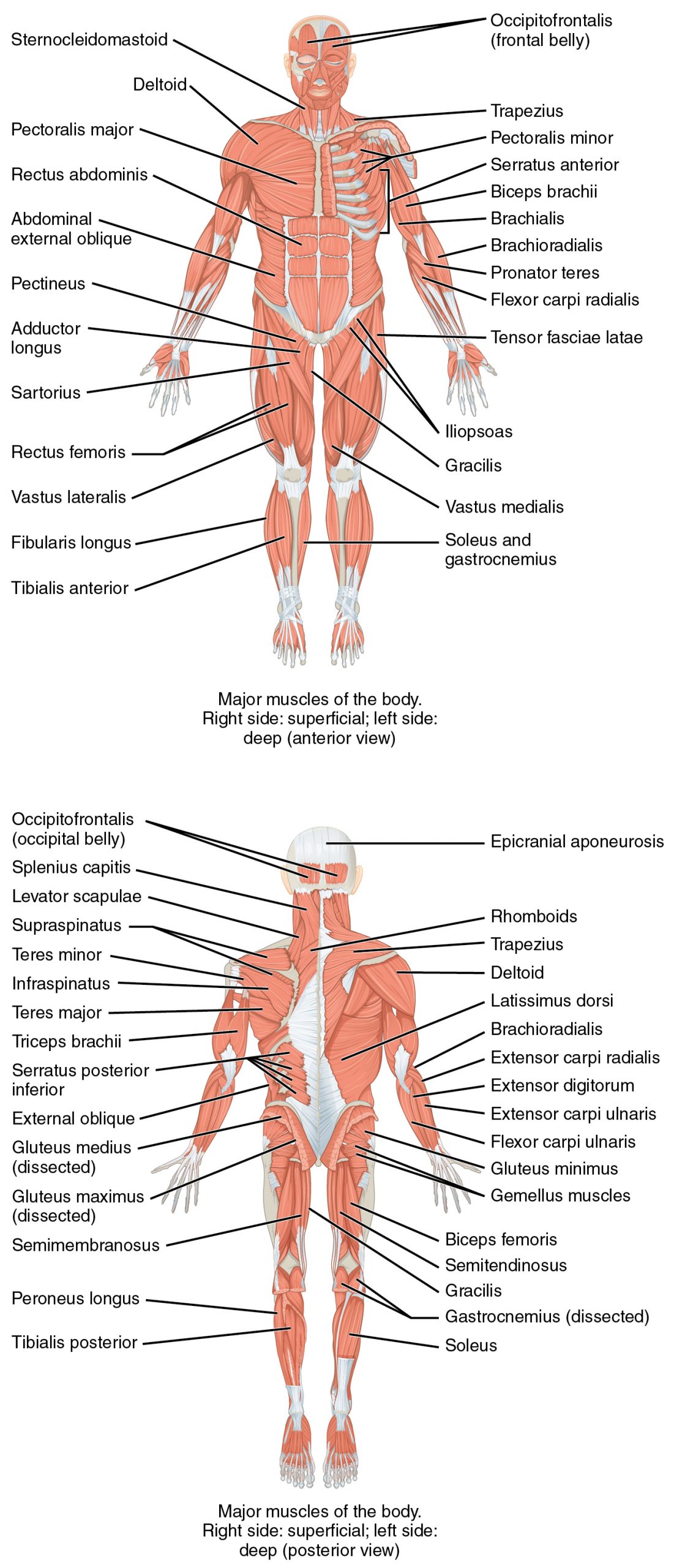
Figure 17.2 image description: The top panel shows the anterior view of the human body with the major muscles labeled. Labels read (from top, head): occipitofrontalis (frontal belly), sternocleidomastoid, trapezius, deltoid, pectorailis minor, serratus anterior, pectoralis major, arm muscles: biceps brachii, brachialis, brachioradialis, pronator teres, flexor carpi radialis, abdomnial: rectus abdominis, abdominal external oblique, lower body: tensor fasciae latae, illiopsoas, penctineus, adductor longus, sartorius, gracilis, rectus femoris, vastus lateralis, vasus medialis, biularis longus, tibialis anterior. The bottom panel shows the posterior view of the human body with the major muscles labeled. Labels read (from top, head, left side): epicranial aponeurosis, occipitofrontalis, splenius capitis, levator scapulae, rhombus, trapezius, supraspinatus, teras minor, infraspinatus, teres major, triceps brachii, seratus posterior inferior, external oblique, lower body: gluteus medius, gluteus maximus, semimebranosus, peroneus longus, tibialis posterior, (right side, from top) trapezius, deltpid, latissimus dorsi, arm: brachioradialis, extersor carpi radialis, extensor digitorum, extensor carpi ulnaris, flexor carpi ulnaris, lower body: gluteus minimus, gemellus muscles, biceps femoris, semitendinosus, gracilis, gastrocnemius, soleus. [Return to Figure 17.2].
Unless otherwise indicated, this chapter contains material adapted from Anatomy and Physiology (on OpenStax), by Betts, et al. and is used under a a CC BY 4.0 international license. Download and access this book for free at https://openstax.org/books/anatomy-and-physiology/pages/1-introduction.
definition
Also known as striated muscles. Skeletal muscles are responsible for voluntary muscle movement.
Is the heart muscle also known as the myocardium. Its appearance is similar to skeletal muscle. It pumps blood and gives the heart beat.
Also known as visceral muscles. Smooth muscle is mainly associated with the walls of internal organs. Smooth muscles are responsible for involuntary muscle movement.
biological process that results in stable equilibrium
in opposition to each other
naming conventions
