4 Integumentary System
Learning Objectives
- Identify general terms related to the integumentary system
- Spell the integumentary system medical terms and use correct abbreviations
- Identify the medical specialties associated with the integumentary system
- Recognize common diseases, disorders, and procedures related to the integumentary system
Chapter Four: Table of Contents
What is it? – General Terms: Integumentary System
What Can Go Wrong? – Diseases, Disorders, and Conditions
- General Conditions, Diseases, and Disorders
- Changes in Skin Color
- Skin Cancer Terminolgy
- Autoimmune Disorders
- Bacterial Infections
- Fungal Infections
- Parasitic Infestations
- Skin Injuries
- Conditions of the Sweat Glands
- Conditions of the Hair and Nails
Integumentary System: General Terms
| Term | Word Breakdown | Description |
|---|---|---|
| integumentary in-tEg-yuh-mEn-tree |
-ary pertaining to integument/o |
The integumentary system forms the barrier between the external environment and the internal systems of the body. In humans, this system consists of skin, hair, nails, and related glands. The main functions are to protect, regulate temperature, and provide sensory function. Read more about the function of the integumentary system |
| cutaneous kyoo-tAY-nee-uhs |
-ous pertaining to cutane/o |
pertaining to the skin (https://www.etymonline.com/word/cutaneous) |
| subcutaneous (subcut) suhb-kyu-tAY-nee-uhs |
-ous pertaining to sub- cutane/o |
Located or placed just beneath the skin. Read about Subcutaneous Injections |
| dermatology duhr-muh-tAH-luh-jee |
-logy study of dermat/o |
the branch of medicine dealing with the skin and its diseases. Read about Dermatology |
| epidermis ep-uh-dUHR-muhs |
dermis -skin epi- |
The outer or top layer of skin, consisting of a layer of dead cells that perform a protective function and a second layer of dividing cells. Read about the function of the epidermis |
| dermis dUHR-muhs |
dermis | The thickest layer of skin located below the epidermis. The dermis contains blood and lymphatic vessels, nerves, glands, and hair follicles. Read more about the dermis. Scroll down the page |
| avascular e-vAs-kyuh-luh |
-ar pertaining to
-a vascul/o |
a lack of blood supply |
| intradermal (ID) in-truh-dUHR-muhl |
-al pertaining to intra- derm/o |
Within the skin. Intradermal injections (ID) are injections administered into the dermis, just below the epidermis. Read about the difference between intradermal (ID) and subcutaneous (subcut) Injections Read about the difference between intradermal (ID) and subcutaneous (subcut) Injections |
| transdermal (TD) trans-dUHR-muhl |
-al pertaining to trans- derm/o |
Through or across the unbroken skin. Read about transdermal patches |
Introduction to the Integumentary System
The integumentary system refers to the skin and its accessory structures. In the adult human body, the skin makes up about 16 percent of body weight and covers an area of 1.5 to 2 m2.
In fact, the skin and accessory structures are the largest organ system in the human body. The skin protects your inner organs and it is in need of daily care and protection to maintain its health.
Watch this video that explains the integumentary system:
Media 6.1. The Integumentary System, 101 Layers of the Skin [Video]. Copyright 2015 by Free School
Integumentary System Word Parts
Click on prefixes, combining forms, and suffixes to reveal a list of word parts to memorize for the Integumentary System.
Practice integumentary system medical terms.
Anatomy (Structures) of the Integumentary System
The skin and its accessory structures make up the integumentary system, which provides the body with overall protection. The skin is made of multiple layers of cells and tissues, which are held to underlying structures by connective tissue. The deeper layer of skin is well vascularized . It also has numerous sensory, and autonomic and sympathetic nerve fibers ensuring communication to and from the brain.
The skin is composed of two main layers:
- The epidermis
- The dermis
- Beneath the dermis lies the hypodermis
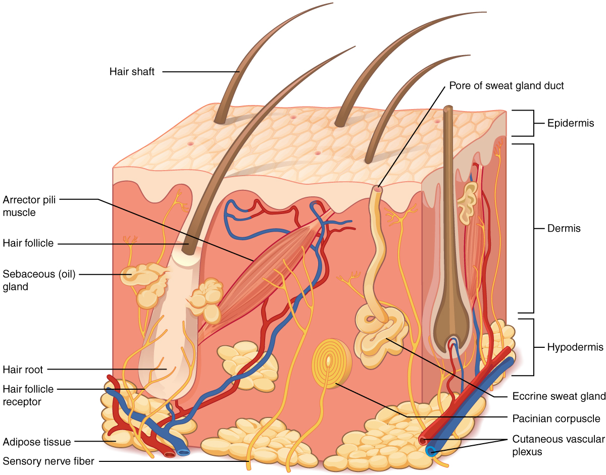
Concept Check
- On the diagram above find the two layers of the skin; epidermis and dermis.
- The literal breakdown for hypodermis is below the dermis. On the diagram above where can you locate it?
- Can you find a hair follicle, hair root and hair shaft?
- Keep reading to find out what the arrector pili muscle does when you are frightened.
Epidermis
The epidermis is composed of keratinized, stratified squamous epithelium. It is made of four or five layers of epithelial cells, depending on its location in the body. It is avascular.
- Thin skin has four layers of cells. From deep to superficial, these layers are the stratum basale, stratum spinosum, stratum granulosum, and stratum corneum. Most of the skin can be classified as thin skin.
- Thick skin is found only on the palms of the hands and the soles of the feet.
The cells in all of the layers except the stratum basale are called keratinocytes. Keratin is an intracellular fibrous protein that gives hair, nails, and skin their hardness and water-resistant properties. The keratinocytes in the stratum corneum are dead and regularly slough away, being replaced by cells from the deeper layers.
Dermis
Hypodermis
The hypodermis serves to connect the skin to the underlying fascia of the bones and muscles. It is not strictly a part of the skin, although the border between the hypodermis and dermis can be difficult to distinguish. The hypodermis consists of well-vascularized, loose, areolar connective tissue and adipose tissue, which functions as a mode of fat storage and provides insulation and cushioning for the integument.
Practice labeling the layers of the skin.
Physiology (Function) of the Integumentary System
The skin and accessory structures perform a variety of essential functions, such as protecting the body from invasion by microorganisms, chemicals, and other environmental factors; preventing dehydration; acting as a sensory organ; modulating body temperature and electrolyte balance; and synthesizing vitamin D. The underlying hypodermis has important roles in storing fats, forming a “cushion” over underlying structures, and providing insulation from cold temperatures.
Protection
The skin protects the body from wind, water, and UV sunlight. It acts as a protective barrier against water loss and it also is the first line of defense against abrasive activity such as grit, microbes, or harmful chemicals. Sweat excreted from sweat glands deters microbes from over-colonizing the skin surface by generating dermicidin, which has antibiotic properties.
Sensory Function
The skin acts as a sense organ because the epidermis, dermis, and the hypodermis contain specialized sensory nerve structures that detect touch, surface temperature, and pain. These receptors are more concentrated on the tips of the fingers, which are most sensitive to touch, especially the Meissner corpuscle, which responds to light touch, and the Pacinian corpuscle , which responds to vibration. Merkel cells, seen scattered in the stratum basale, are also touch receptors. In addition to these specialized receptors, there are sensory nerves connected to each hair follicle, pain and temperature receptors scattered throughout the skin, and motor nerves innervate the arrector pili muscles and glands. This rich innervation helps us sense our environment and react accordingly,
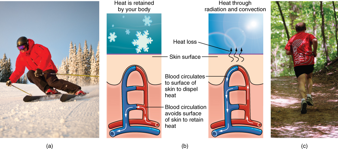
Thermoregulation
The integumentary system helps regulate body temperature through its tight association with the sympathetic nervous system. The sympathetic nervous system is continuously monitoring body temperature and initiating appropriate motor responses.
- When the body becomes warm sweat glands, accessory structures to the skin, secrete water, salt, and other substances to cool the body.
- Even when the body does not appear to be noticeably sweating, approximately 500 mL of sweat are secreted a day.
- If the body becomes excessively warm due to high temperatures, vigorous activity, or a combination of the two, sweat glands will be stimulated by the sympathetic nervous system to produce large amounts of sweat.
- When the sweat evaporates from the skin surface, the body is cooled as body heat is dissipated.
- In addition to sweating, arterioles in the dermis dilate so that excess heat carried by the blood can dissipate through the skin and into the surrounding environment (Figure 2b).
- This accounts for the skin redness that many people experience when exercising.
- When body temperatures drop, the arterioles constrict to minimize heat loss, particularly in the ends of the digits and tip of the nose.
- This reduced circulation can result in the skin taking on a whitish hue.
- Although the temperature of the skin drops as a result, passive heat loss is prevented, and internal organs and structures remain warm.
- If the temperature of the skin drops too much (such as environmental temperatures below freezing), the conservation of body core heat can result frostbite .
Concept Check
Can you describe the thermoregulation process between the integumentary system and the sympathetic system?
- When body temperature is too warm.
- When body temperature is too cold.
Accessory Structures
Accessory structures of the skin include hair, nails, sweat glands, and sebaceous glands. These structures embryologically originate from the epidermis and can extend down through the dermis into the hypodermis.
Hair
Hair is a keratinous filament growing out of the epidermis. It is primarily made of dead, keratinized cells. Strands of hair originate in an epidermal penetration of the dermis called the hair follicle. The hair shaft is the part of the hair not anchored to the follicle, and much of this is exposed at the skin’s surface. The rest of the hair, which is anchored in the follicle, lies below the surface of the skin and is referred to as the hair root. The hair root ends deep in the dermis at the hair bulb, and includes a layer of mitotically active basal cells called the hair matrix. The hair bulb surrounds the hair papilla, which is made of connective tissue and contains blood capillaries and nerve endings from the dermis.
Hair Function
Hair serves a variety of functions, including protection, sensory input, thermoregulation, and communication. For example:
- Hair on the head protects the skull from the sun.
- Hair in the nose and ears, and around the eyes (eyelashes) defends the body by trapping and excluding dust particles that may contain allergens and microbes.
- Hair of the eyebrows prevents sweat and other particles from dripping into and bothering the eyes.
Hair also has a sensory function due to sensory innervation by a hair root plexus surrounding the base of each hair follicle. Hair is extremely sensitive to air movement or other disturbances in the environment, much more so than the skin surface. This feature is also useful for the detection of the presence of insects or other potentially damaging substances on the skin surface.
Each hair root is connected to a smooth muscle called the arrector pili that contracts in response to nerve signals from the sympathetic nervous system, making the external hair shaft “stand up.” The primary purpose for this is to trap a layer of air to add insulation. This is visible in humans as goose bumps and even more obvious in animals, such as when a frightened cat raises its fur. Of course, this is much more obvious in organisms with a heavier coat than most humans, such as dogs and cats.
Hair Growth, Loss and Colour
Hair grows and is eventually shed and replaced by new hair. Hair typically grows at the rate of 0.3 mm per day. On average, 50 hairs are lost and replaced per day. Hair loss occurs if there is more hair shed than what is replaced and can happen due to hormonal or dietary changes. Hair loss can also result from the aging process, or the influence of hormones. Similar to the skin, hair gets its colour from the pigment melanin, produced by melanocytes in the hair papilla. Different hair color results from differences in the type of melanin. As a person ages, the melanin production decreases, and hair tends to lose its color and becomes gray and/or white.
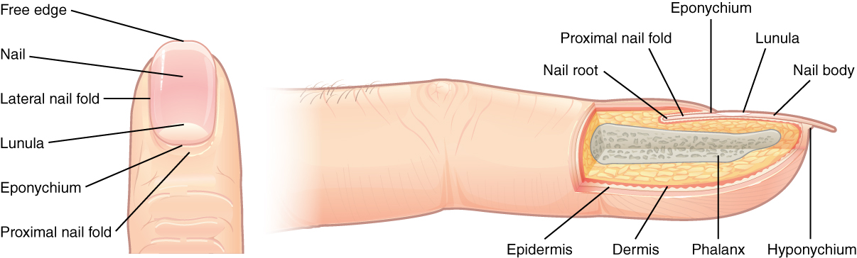
Nails
The nail bed is a specialized structure of the epidermis that is found at the tips of our fingers and toes. The nail body is formed on the nail bed, and protects the tips of our fingers and toes as they are the farthest extremities and the parts of the body that experience the maximum mechanical stress (see Figure 6.3). The nail body forms a back-support for picking up small objects with the fingers. The nail body is composed of densely packed dead keratinocytes.
The epidermis in this part of the body has evolved a specialized structure upon which nails can form. The nail body forms at the nail root, which has a matrix of proliferating cells from the stratum basale that enables the nail to grow continuously. The lateral nail fold overlaps the nail on the sides, helping to anchor the nail body. The nail fold that meets the proximal end of the nail body forms the nail cuticle, also called the eponychium.
The nail bed is rich in blood vessels, making it appear pink, except at the base, where a thick layer of epithelium over the nail matrix forms a crescent-shaped region called the lunula (the “little moon”). The area beneath the free edge of the nail, furthest from the cuticle, is called the hyponychium. It consists of a thickened layer of stratum corneum.
Sweat Glands
Sudoriferous Glands
When the body becomes warm, sudoriferous glands produce sweat to cool the body.
Sebaceous Glands
A sebaceous gland is a type of oil gland that is found all over the body and helps to lubricate and waterproof the skin and hair. Most sebaceous glands are associated with hair follicles. They generate and excrete sebum, a mixture of lipids, onto the skin surface, thereby naturally lubricating the dry and dead layer of keratinized cells of the stratum corneum, keeping it pliable. The fatty acids of sebum also have antibacterial properties, and prevent water loss from the skin in low-humidity environments. The secretion of sebum is stimulated by hormones, many of which do not become active until puberty. Thus, sebaceous glands are relatively inactive during childhood.
Words not Easily Broken into Word Parts
Just a Little Practice!
Changes Due to Aging
All systems in the body accumulate subtle and some not-so-subtle changes as a person ages. Among these changes are reductions in cell division, metabolic activity, blood circulation, hormonal levels, and muscle strength (see Figure 6.4). In the skin, these changes are reflected in decreased mitosis in the stratum basale, leading to a thinner epidermis. The dermis, which is responsible for the elasticity and resilience of the skin, exhibits a reduced ability to regenerate, which leads to slower wound healing. The hypodermis, with its fat stores, loses structure due to the reduction and redistribution of fat, which in turn contributes to the thinning and sagging of skin.
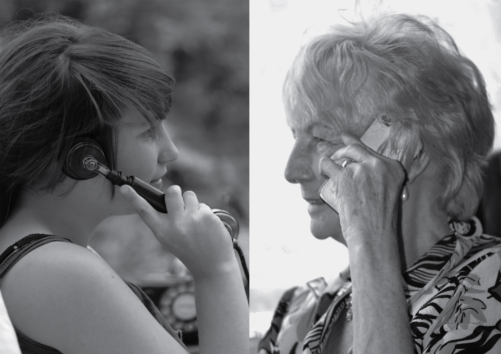
The accessory structures also have lowered activity, generating thinner hair and nails, and reduced amounts of sebum and sweat. A reduced sweating ability can cause some elderly to be intolerant to extreme heat. Other cells in the skin, such as melanocytes and dendritic cells, also become less active, leading to a paler skin tone and lowered immunity. Wrinkling of the skin occurs due to breakdown of its structure, which results from decreased collagen and elastin production in the dermis, weakening of muscles lying under the skin, and the inability of the skin to retain adequate moisture.
The integumentary system is susceptible to a variety of diseases, conditions, and injuries. These range from annoying but relatively benign bacterial or fungal infections that are categorized as disorders, to skin cancer and severe burns, which can be fatal. In this section, you will learn several of the most common skin conditions.
General Diseases, Disorders and Conditions
| Term | Word Breakdown | Description |
|---|---|---|
| dermatitis duhr-muh-tIE-tuhs |
-itis inflammation dermat/o |
A general term for conditions that cause inflammation of the skin. (https://my.clevelandclinic.org/health/articles/4089-dermatitis) |
| eczema Eg-zuh-muh |
A common skin condition that causes itchiness, rashes, dry patches, and infection. It is not contagious Read more about eczema | |
| petechiae puh-tEE-kee-ie |
Tiny red, flat spots that appear on your skin. Petechiae are a sign of blood leaking from capillaries under your skin. Read more about petechiae | |
| contusion kuhn-tOO-zhuhn |
Another word for bruise. A contusion (bruise) is an injury to the soft tissue often produced by a blunt force such as a kick, fall, or blow. It does not break the skin. The immediate result will be pain, swelling, and discoloration (https://stanfordhealthcare.org/medical-conditions/bones-joints-and-muscles/sports-related-injuries/types/contusion.html). | |
| ecchymosis ek-i-mOH-suhs |
-osis condition |
A type of bruise where the mark is a 1/2 inch long or bigger. Read more about ecchymosis |
| hemaoma hee-muh-tOH-muht |
-oma tumor; mass; fluid collection hemat/o |
An abnormal collection of blood outside of a blood vessel. It occurs because the wall of a blood vessel wall, artery, vein, or capillary, has been damaged and blood has leaked into tissues where it does not belong. Hematomas may cause symptoms of inflammation including pain, swelling, and redness. Read more about hematomas |
| necrosis nuh-krOH-suhs |
-osis condition necr/o |
The death of body tissue. It occurs when too little blood flows to the tissue. This can be from injury, radiation, or chemicals. Necrosis cannot be reversed. (Medline Plus) |
| pruritus proo-rIE-tuhs |
itis- inflammation prurit/o |
Itching. Pruritus is symptom for other conditions. Read more about pruritus |
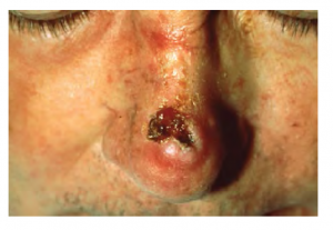
| rhinophyma | -phý̄ma a swelling, tumor, equivalent. rhin/o |
A skin disorder affecting your nose. It causes your nose to grow larger and appear red, bumpy, and rounded. Read more about rhinophyma |
| xeroderma | -derma skin Combining form |
Dry skin (xeroderma. (n.d.) American Heritage® Dictionary of the English Language, Fifth Edition. (2011). Retrieved August 19 2023 from https://www.thefreedictionary.com/xeroderma) |
| hemangioma hee man jee OH mah |
-oma tumor; mass; fluid collection hem/o angi/o |
A common vascular birthmark, made of extra blood vessels in the skin. It is a benign (non-cancerous) growth. Read more about hemangiomas |
| papilloma pap-uh-lOH-muh |
-oma tumor; mass; fluid collection papill/o |
A small wart like growth on the skin or on a mucous membrane, derived from the epidermis and usually benign. Read more about papillomas |
| syndactyly sin-dAk-tuh-lee |
syn: together
dactyl/o |
Fingers or toes (digits) may be fused together (syndactyly) webbed feet or hands. See images of and read more about syndactly |
| polydactyly pah-lee-dAk -tuh-lee |
-y noun ending poly- dactyl/o |
Extra fingers or toes |
| verruca vuhr-rOO-kuh |
Warts; warts an be viral or fungal. Read about types of warts |
Eczema
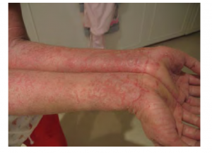
A common skin disorder is eczema Eczema is an inflammatory condition and occurs in individuals of all ages. Acne involves the clogging of pores, which can lead to infection and inflammation, and is often seen in adolescents. Other disorders, include seborrheic dermatitis (on the scalp), psoriasis, fungal infections, cold sores, impetigo, scabies, hives, and warts (Betts, et al., 2013).
Eczema is an allergic reaction that manifests as dry, itchy patches of skin that resemble rashes (see Figure 6.5). It may be accompanied by swelling of the skin, flaking, and in severe cases, bleeding. Symptoms are usually managed with moisturizers, corticosteroid creams, and immunosuppressants (Betts, et al., 2013).
Pop Quiz Time!
Changes in Skin Color
| Term | Word Breakdown | Description |
|---|---|---|
| albinism Al-buh-niz-uhm |
-ism condition albin/o |
Born with little or no pigment in your hair, eyes, and skin. The missing pigment is called melanin. Albinism usually makes your coloring lighter than is typical for your family or ethnic background. Read more about albinism |
| cyanosis sie-uh-nOH-suhs |
-osis condition cyan/o |
Bluish discoloration of the skin usually due to a lack of oxygen in the blood. Read more about cyanosis |
| erythroderma | -a noun ending erythr- derm/o |
Reddening of the skin. Read more about erythroderma |
| leukoderma | -a noun ending leuk- derm/o |
A clinical sign describing a localized area of white spots on the skin. Usually has an accidental cause. Read more about leukoderma |
| vitiligo vit-uh-lIE-goh |
A long-term condition where pale white patches develop on the skin. It's caused by the lack of melanin, which is the pigment in skin. Often due to an autoimmune response. |
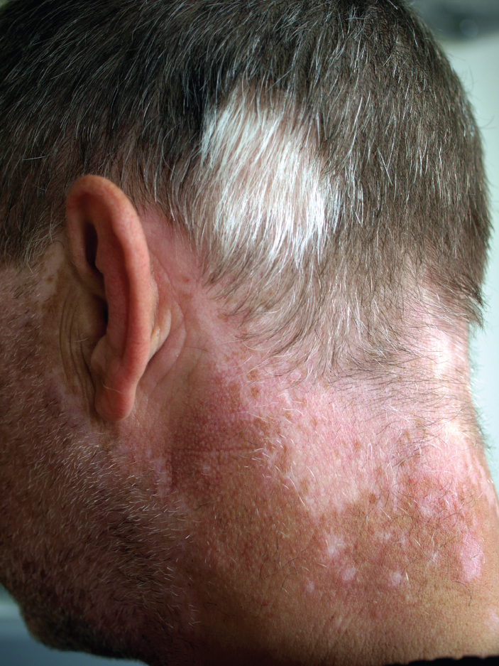
Some disorders are marked by changes in skin color.
Albinism
Albinism is a genetic disorder that affects (completely or partially) the coloring of skin, hair, and eyes. This is primarily due to the inability of melanocytes to produce melanin. Individuals with albinism tend to appear white or very pale due to the lack of melanin in their skin and hair. Recall that melanin helps protect the skin from the harmful effects of UV radiation. Individuals with albinism tend to need more protection from UV radiation, as they are more prone to sunburns and skin cancer. They also tend to be more sensitive to light and have vision problems due to the lack of pigmentation on the retinal wall (Betts, et al., 2013)
Treatment of this disorder usually involves addressing the symptoms, such as limiting UV light exposure to the skin and eyes. In vitiligo, the melanocytes in certain areas lose their ability to produce melanin, possibly due to an autoimmune reaction. This leads to a loss of color in patches (see Figure 6.6). Neither albinism nor vitiligo directly affects the lifespan of an individual (Betts, et al., 2013)
Cyanosis
A prolonged reduction in oxygen levels, dark red deoxyhemoglobin becomes dominant in the blood, making the skin appear blue, a condition referred to as cyanosis. This happens when the oxygen supply is restricted, as when someone is experiencing difficulty in breathing because of asthma or a heart attack. However, in these cases the effect on skin color has nothing do with the skin’s pigmentation (Betts, et al., 2013)
Test Your Knowledge
Skin Cancer Terminology
| Term | Word Breakdown | Description |
|---|---|---|
| benign (beh-NINE) |
Not cancerous does not spread. | |
| metastasize muh-ta-stuh-size |
Cancer that spreads | |
| neoplasm NEE-oh-PLA-zum |
-ous pertaining to sub cutane/o |
New growth distinct from the tissue in which it occurs, May or may not be cancerous. Read more about neoplasms |
| basal cell carcinoma (BCC) bAY-suhl sEl kahr-suh-nOH-muh |
-al pertaining to bas/o -oma carcin/o |
The most common type of skin cancer. Basal cell carcinoma begins in the basal cells of the epidermis. It grows slowly and rarely spreads to other body parts. Read more about basal cell carcinomas (bcc) |
| malignant melanoma muh-lIg-nuhnt mel-uh-nOH-muh |
-oma tumor; mass; fluid collection melan/o - |
A melanoma is a cancer characterized by the uncontrolled growth of melanocytes, the pigment-producing cells in the epidermis. Read more about melanoma |
| squamous cell carcinoma (SCC) skwAY-muhs sEl kahr-suh-nOH-muh |
-oma tumor; mass; fluid collections \carcin/o - cancer |
A cancer that affects the keratinocytes of the stratum spinosum and presents as lesions commonly found on the scalp, ears, and hands Read more about squamous cell carcinomas (SCC) Read more about squamous cell carcinomas (SCC) |
One of the most talked about diseases is skin cancer. Most cancers are identified by the organ or tissue in which the cancer originates. One common form of cancer is skin cancer.
In general, cancers result from an accumulation of DNA mutations. These mutations can result in cell populations that do not die when they should and uncontrolled cell proliferation that leads to tumors. Although many tumors are benign, some metastasize. Cancers are characterized by their ability to metastasize.
Sun Damage
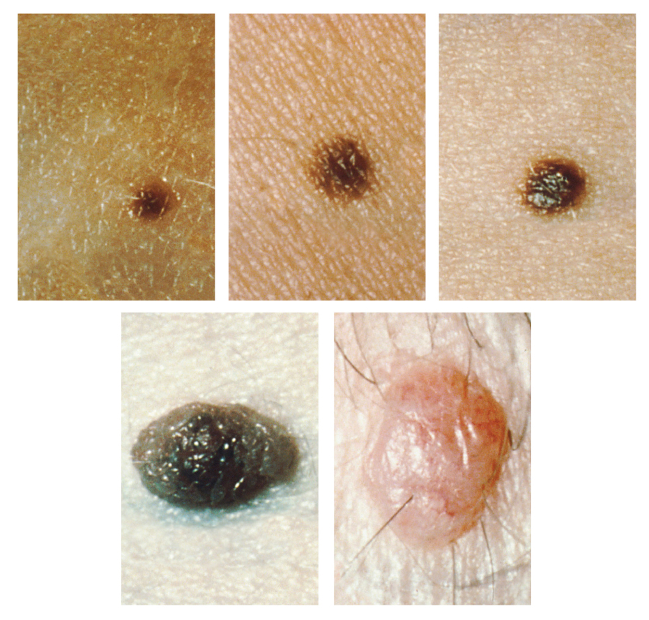
It requires about 10 days after initial sun exposure for melanin synthesis to peak, which is why pale-skinned individuals tend to suffer sunburns of the epidermis initially. Dark-skinned individuals can also get sunburns, but are more protected than are pale-skinned individuals. Too much sun exposure can eventually lead to wrinkling due to the destruction of the cellular structure of the skin, and in severe cases, can cause sufficient DNA damage to result in skin cancer. When there is an irregular accumulation of melanocytes in the skin, freckles appear. Moles are larger masses of melanocytes, and although most are benign, they should be monitored for changes that might indicate the presence of cancer (see Figure 6.7).
Basal Cell Carcinoma (BCC)
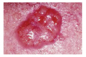
Basal cell carcinoma is a form of cancer that affects the mitotically active stem cells in the stratum basale of the epidermis. It is the most common of all cancers that occur in the United States and is frequently found on the head, neck, arms, and back, which are are as that are most susceptible to long-term sun exposure. Although UV rays are the main culprit, exposure to other agents, such as radiation and arsenic, can also lead to this type of cancer. Wounds on the skin due to open sores, tattoos, burns, etc. may be predisposing factors. Basal cell carcinomas start in the stratum basale and usually spread along this boundary. At some point, they begin to grow toward the surface and become an uneven patch, bump, growth, or scar on the skin surface (see Figure 6.8). Like most cancers, basal cell carcinomas respond best to treatment when caught early. Treatment options include surgery, freezing (cryosurgery), and topical ointments.
Squamous Cell Carcinoma (SCC)
Squamous cell carcinoma is a cancer that affects the keratinocytes of the stratum spinosum and presents as lesions commonly found on the scalp, ears, and hands (see Figure 6.9). It is the second most common skin cancer. The American Cancer Society reports that two of 10 skin cancers are squamous cell carcinomas, and it is more aggressive than basal cell carcinoma. If not removed, these carcinomas can metastasize. Surgery and radiation are used to cure squamous cell carcinoma.
Melanoma
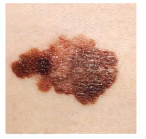
A melanoma is a cancer characterized by the uncontrolled growth of melanocytes, the pigment-producing cells in the epidermis. Typically, a melanoma develops from a mole. It is the most fatal of all skin cancers, as it is highly metastatic and can be difficult to detect before it has spread to other organs. Melanomas usually appear as asymmetrical brown and black patches with uneven borders and a raised surface (see Figure 6.10). Treatment typically involves surgical excision and immunotherapy.
ABCDE for Early Diagnosis
Doctors often give their patients the following ABCDE mnemonic to help with the diagnosis of early-stage melanoma. If you observe a mole on your body displaying these signs, consult a doctor.
Asymmetry – the two sides are not symmetrical
Borders – the edges are irregular in shape
Color – the color is varied shades of brown or black
Diameter – it is larger than 6 mm (0.24 in)
Evolving – its shape has changed
Some specialists cite the following additional signs for the most serious form, nodular melanoma:
Elevated – it is raised on the skin surface
Firm – it feels hard to the touch
Growing – it is getting larger
Know your Skin Cancer!
Autoimmune Disorders
| Term | Word Breakdown | Description |
|---|---|---|
| psoriasis suhr-rIE-uh-suhs |
-iasis condition, formation of psor/o |
A skin disease marked by red, itchy, scaly patches. |
| scleroderma (sklair-oh-DUR-muh) |
-derma skin scler/o |
A type of autoimmune disorder. In this condition, the immune system mistakenly attacks and damages healthy body tissue. It leads to hard thickened skin. (https://medlineplus.gov/ency/article/000429.htm) |
Autoimmune system disorders cause an over activity of the immune system. The the body attacks and damages its own tissues . (Web MD).
Psoriasis
Psoriasis is a chronic autoimmune disorder that results in patches of thick red skin with the appearance of silvery scales. These patches can be found on elbows, knees, scalp, low back, face, feet, fingernails, toenails and even the mouth. Psoriasis can be confused with other skin disease so a dermatologist is the best physician to diagnosis psoriasis. Treatments may include creams, ointments, ultraviolet light therapy and medication (Center for Disease Control and Prevention, 2018). To learn more, visit the Center for Disease Control and Prevention’s web page on psoriasis.
Autoimmune Disease Pop Quiz
Bacterial Infections
| Term | Word Breakdown | Description |
|---|---|---|
| abscess Ab-ses |
A localized collection of pus, usually caused by an infection. (https://medlineplus.gov/abscess.html) | |
| acne Ak-nee |
A skin condition that occurs when your pores become plugged with oil and dead skin cells. It causes pimples, blackheads, and whiteheads. (https://www.mayoclinic.org/diseases-conditions/acne/symptoms-causes/syc-20368047). | |
| cellulitis sel-yuh-lIE-tuhs |
A common bacterial skin infection that causes redness, swelling, and pain in the infected area of the deeper layers of the skin. If untreated, it can spread and cause serious health problems.
Good wound care and hygiene are important for preventing cellulitis. |
|
| folliculitis fuh-lik-yuh-lIE-tuhs |
-itis inflammation follicul/o |
inflammation of the hair follicles, usually caused by infection or irritation (https://www.webmd.com/skin-problems-and-treatments/what-is-folliculitis) |
| impetigo im-puh-TIE-go |
A common and highly contagious skin infection that mainly affects infants and young children. Bacteria infects the skin, they cause sores. Symptoms include red, itchy sores that break open and leak a clear fluid or pus for a few days. The bacteria can spread to others if someone touches those sores or comes into contact with fluid from the sores.(https://www.cdc.gov/groupastrep/diseases-public/impetigo.html) |
Acne
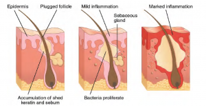
Acne is a skin disturbance that typically occurs on areas of the skin that are rich in sebaceous glands (face and back). It is most common along with the onset of puberty due to associated hormonal changes, but can also occur in infants and continue into adulthood. Hormones, such as androgens, stimulate the release of sebum. An overproduction and accumulation of sebum along with keratin can block hair follicles. This plug is initially white. The sebum, when oxidized by exposure to air, turns black. Acne results from infection by acne-causing bacteria (Propionibacterium and Staphylococcus), which can lead to redness and potential scarring due to the natural wound healing process (see Figure 6.11) (Betts, et al., 2013).
Bacterial Infection? Who Am I!
Fungal Infections
Ringworm
Tinea or dermatophytosis dermat/o/phyt/o/sis often referred to as ringworm. The name ringworm presents as a circular rash that is itchy and red and can be found on various parts of the body. It is referred to by the location that it is found:
| Term | Word Breakdown | Description |
|---|---|---|
| dermatophytosis | -osis
dermat/o - Combining form: phyt/o |
A common fungal infection of the skin and nails that is caused by fungus known as dermatophytes. Dermatophytes belong to a group of organisms that are able to break down the keratin in tissues such as the epidermis, hair, nails, feathers, horns and hooves.(https://www.cfsph.iastate.edu/Factsheets/pdfs/dermatophytosis.pdf) Also known as ringworm. |
| tinea capitus tIn-ee-uh |
Fungal infection or ringworm of the scalp | |
| tinea corporis tIn-ee-uh |
corpor/o body |
Fungal infection or ringworm of the scalp |
| tinea pedis tIn-ee-uh |
ped/o child; foot |
Fungal infection or ringworm of the feet. commonly referred to as athlete's foot |
| tinea unguium tIn-ee-uh |
-um structure ungu/o |
Fungal infection or ringworm of the toenails and fingernails. Also called onychomycosis |
“Fun Guy” Quiz!
To learn more about ringworm, visit the Center for Disease Control and Prevention’s web page on fungal infections.
Parasitic Infestations
As the skin is constantly exposed to the environment, it is a common portal of entry for parasites. Infestations with parasites can cause systemic diseases in humans but often result in cutaneous lesions which are on the rise in dermatology (News Medical Life Sciences)
| Term | Word Breakdown | Description |
|---|---|---|
| pediculosis pi-dik-yuh-lOH-suhs |
-osis condition pedicul/o |
Lice |
| scabies skAY-beez |
Human scabies is caused by an infestation of the skin by the human itch mite. It can be transmitted through skin-to-skin contact.(https://www.cdc.gov/parasites/scabies/index.html#:~:text=Parasites%20%2D%20Scabies,-Print&text=Human%20scabies%20is%20caused%20by,a%20pimple%2Dlike%20skin%20rash.) |
Parasite, Para bite!
Skin Injuries
Integumentary System: Terms that Describe Skin Injuries
| Term | Word Breakdown | Description |
|---|---|---|
| abrasion uh-brAY-zhuhn |
An abrasion is a scrape or graze of the skin. An abrasion can happen to a person, as in a skinned knee, or to an object, as in what you get when you apply sandpaper to wood. ("Abrasion." Vocabulary.com Dictionary, Vocabulary.com, https://www.vocabulary.com/dictionary/abrasion. | |
| blister blIs-tuhr |
A blister is a fluid-filled bubble that forms on the upper layers of the skin after rubs or burns on the skin (Vocabulary.com. (n.d.). Blister. In Vocabulary.com Dictionary. Retrieved August 22, 2023, from https://www.vocabulary.com/dictionary/blister) | |
| callus kAl-uhs |
A callus is a spot where your skin becomes rough and thick after repeated pressure or friction. (Vocabulary.com. (n.d.). Callus. In Vocabulary.com Dictionary. Retrieved August 22, 2023, from https://www.vocabulary.com/dictionary/callus | |
| decubitus ulcer UHl-suhr |
Decubitus ulcers, also termed bedsores or pressure ulcers, are skin and soft tissue injuries that form as a result of constant or prolonged pressure exerted on the skin. These ulcers occur at bony areas of the body. They come when people are immobile and lay in one area for too long (https://www.ncbi.nlm.nih.gov/books/NBK553107/#:~:text=Decubitus%20ulcers%2C%20also%20termed%20bedsores,than%20medial)%2C%20and%20occiput.) | |
| keloid kEE-loid |
-oid
kel/o |
Overgrowth of scar tissue (https://www.mayoclinic.org/diseases-conditions/keloid-scar/symptoms-causes/syc-20520901) |
Skin injuries set off a healing process that occurs in several overlapping stages.
- The first step to repairing damaged skin is the formation of a blood clot that helps stop the flow of blood and scabs over with time. Many different types of cells are involved in wound repair, especially if the surface area that needs repair is extensive.
- Before the basal stem cells of the stratum basale can recreate the epidermis, fibroblasts mobilize and divide rapidly to repair the damaged tissue by collagen deposition, forming granulation tissue.
- Blood capillaries follow the fibroblasts and help increase blood circulation and oxygen supply to the area.
- Immune cells, such as macrophages, roam the area and engulf any foreign matter to reduce the chance of infection (Betts, et al., 2013).
Scars and Keloids
Most cuts or wounds, with the exception of ones that only scratch the epidermis, lead to scar formation. Scarring occurs in cases in which there is repair of skin damage, but the skin fails to regenerate the original skin structure. Fibroblasts generate scar tissue in the form of collagen, and the bulk of repair is due to the basket-weave pattern generated by collagen fibers and does not result in regeneration of the typical cellular structure of skin. Instead, the tissue is fibrous in nature and does not allow for the regeneration of accessory structures, such as hair follicles, sweat glands, or sebaceous glands (Betts, et al., 2013).
Sometimes, there is an overproduction of scar tissue, because the process of collagen formation does not stop when the wound is healed; this results in a keloid. In contrast, scars that result from acne and chickenpox have a sunken appearance and are called atrophic scars (Betts, et al., 2013)
Scarring of skin after wound healing is a natural process and does not need to be treated further. Application of mineral oil and lotions may reduce the formation of scar tissue. However, modern cosmetic procedures, such as dermabrasion, laser treatments, and filler injections have been invented as remedies for severe scarring. All of these procedures try to reorganize the structure of the epidermis and underlying collagen tissue to make it look more natural (Betts, et al., 2013).
Bedsores and Stretch Marks
Skin and its underlying tissue can be affected by excessive pressure. One example of this is called a bedsore. Bedsores, also called decubitus ulcers, are caused by constant, long-term, unrelieved pressure on certain body parts that are bony, reducing blood flow to the area and leading to necrosis . Bedsores are most common in elderly patients who have debilitating conditions that cause them to be immobile. Most hospitals and long-term care facilities have the practice of turning the patients every few hours to prevent the incidence of bedsores. If left untreated bedsores can be fatal if they become infected (Betts, et al., 2013)
The skin can also be affected by pressure associated with rapid growth. A stretch mark results when the dermis is stretched beyond its limits of elasticity, as the skin stretches to accommodate the excess pressure. Stretch marks usually accompany rapid weight gain during puberty and pregnancy. They initially have a reddish hue, but lighten over time. Other than for cosmetic reasons, treatment of stretch marks is not required. They occur most commonly over the hips and abdomen (Betts, et al., 2013).
Calluses
When you wear shoes that do not fit well and are a constant source of abrasion on your toes, you tend to form a callus at the point of contact. This occurs because the basal stem cells in the stratum basale are triggered to divide more often to increase the thickness of the skin at the point of abrasion to protect the rest of the body from further damage. This is an example of a minor or local injury, and the skin manages to react and treat the problem independent of the rest of the body. Calluses can also form on your fingers if they are subject to constant mechanical stress, such as long periods of writing, playing string instruments, or video games. A corn is a specialized form of callus. Corns form from abrasions on the skin that result from an elliptical-type motion (Betts, et al., 2013).
Burns
A burn results when the skin is damaged by intense heat, radiation, electricity, or chemicals. The damage results in the death of skin cells, which can lead to a massive loss of fluid. Dehydration, electrolyte imbalance, and renal and circulatory failure follow, which can be fatal. Burn patients are treated with intravenous fluids to offset dehydration, as well as intravenous nutrients that enable the body to repair tissues and replace lost proteins. Another serious threat to the lives of burn patients is infection. Burned skin is extremely susceptible to bacteria and other pathogens, due to the loss of protection by intact layers of skin (Betts, et al., 2013).
Burn Classification
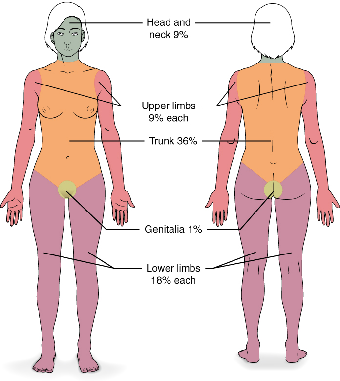
Burns are sometimes measured in terms of the size of the total surface area affected. This is referred to as the rule of nines, which associates specific anatomical areas with a percentage that is a factor of nine (see Figure 6.12) (Betts, et al., 2013).
Burns are also classified by the degree of their severity.
- A first-degree burn is a superficial burn that affects only the epidermis. Although the skin may be painful and swollen, these burns typically heal on their own within a few days. Mild sunburn fits into the category of a first-degree burn.
- A second-degree burn goes deeper and affects both the epidermis and a portion of the dermis. These burns result in swelling and a painful blistering of the skin. It is important to keep the burn site clean and sterile to prevent infection. If this is done, the burn will heal within several weeks.
- A third-degree burn fully extends into the epidermis and dermis, destroying the tissue and affecting the nerve endings and sensory function. These are serious burns that may appear white, red, or black; they require medical attention and will heal slowly without it.
- A fourth-degree burn is even more severe, affecting the underlying muscle and bone.
Oddly, third and fourth-degree burns are usually not as painful because the nerve endings themselves are damaged. Full-thickness burns cannot be repaired by the body, because the local tissues used for repair are damaged and require debridement, or amputation in severe cases, followed by grafting of the skin from an unaffected part of the body, or from skin grown in tissue culture for grafting purposes. Skin grafts are required when the damage from trauma or infection cannot be closed with sutures or staples (Betts et al., 2013).
Skin Injuries Activity
Conditions of the Sweat Glands
| Term | Word Breakdown | Description |
|---|---|---|
| anhidrosis | -osis condition -an hidr/o |
Not being able to sweat. normally |
| diaphoresis die-uh-fuhr-rEE-suhs |
-esis state or condition diaphor/o phor/o |
Excessive sweating without an obvious cause, such as heat or exercise (https://www.medicalnewstoday.com/articles/321663) |
Sweaty Conditions
Conditions of the Hair and Nails
| Term | Word Breakdown | Description |
|---|---|---|
| alopecia al-uh-pEE-shuh |
baldness | |
| hirsutism hUHR-suh-tiz-uhm |
Condition where women have thick, dark hair on their face, neck, chest, tummy, lower back, buttocks or thighs (https://www.nhs.uk/conditions/hirsutism/) | |
| onychomycosis | -osis condition onych/o myc/o |
nail fungus |
Medical and Surgical Procedures
| Term | Word Breakdown | Description |
|---|---|---|
| allograft Al-uh-graft |
-graft any tissue or organ for implantation or transplantation allo/o |
Reconstructive surgery where tissue or an organ is taken from a donor. Read more about the pros and cons of allografts and autografts |
| autograft AW-toh-graft |
-graft any tissue or organ for implantation or transplantation aut/o |
Reconstructive surgery where tissue is removed from one part of the body and grafted on to another part of the body (same person). Read more about the pros and cons of allografts and autografts |
| biopsy (Bx) bIE-ahp-see |
-opsy visual examination bi/o |
A biopsy is a medical procedure that involves taking a small sample of body tissue so it can be examined under a microscope. Read more about biopsies |
| cryosurgery krie-oh-sUHRj-ree |
Procedure that uses freezing cold temperatures to kill abnormal cells or small tumors. Read more about cryosurgery | |
| debridement di-brEEd-muhnt |
Debridement is the removal of dead (necrotic) or infected skin tissue to help a wound heal. It’s also done to remove foreign material from tissue. Read more about debridement | |
| dermabrasion duhr-muh-brAY-zhuhn |
-ion process derm/o |
A procedure used to exfoliate the outer layer of the skin. Read more about dermabrasion |
| dermatoplasty | - plasty surgical reconstruction dermat/o |
Plastic surgery or surgical reconstruction of the skin. |
| xenograft zEn-uh-graft |
-graft any tissue or organ for implantation or transplantation xen/o |
A graft in which the donor and recipient are of different species. (https://www.online-dictionary.com/what-are/the-definitions-of/xenograft) |
Procedure Pop Quiz
Drug Categories
| Term | Word Breakdown | Description |
|---|---|---|
| anesthetic an-uhs-thEt-ik |
-ic pertaining to an- |
Type of drug that removes feeling or sensation |
| antifungal an-tee-fUHng-guhl |
-al pertaining to anti- fung/o |
Medication used to treat fungus. |
| antipruritic an-tee-proo-rIt-ik |
-ic pertaining to anti- prurit/o |
Drug used to reduce itching. |
| corticosteroid kor-tik-oh-stIR-oid |
-oid resembling ster/o |
Drug that reduces inflammation |
Know your Drugs!
Careers
Integumentary System: Career Focus
| Term | Word Breakdown | Description |
|---|---|---|
| dermatologist dur-muh-tol-uh-jist |
Suffix: -logist one who studies Combining form: dermat/o skin |
A dermatologist is a medical doctor with specialized training in treating diseases, disorders and injuries related to the integumentary system and its accessory structures. |
| esthetician | Suffix: -ian having a certain profession. Combining form: |
An esthetician is a trained technician who specializes in skin beautification. |
| plastic surgeon | suffix: -ic pertaining to Combining form: |
A dermatologist is a medical doctor with specialized training in treating diseases, disorders and injuries related to the integumentary system and its accessory structures. There are many dermatologic subspecialties such as cosmetic dermatology, dermatopathology and pediatric dermatology. To learn more visit the Dermatology and Subspecialties section of the Canadian Dermatology Association website.
Dermatologists can be specially trained to perform a procedure called Mohs surgery. Mohs surgery excises skin cancers in thin layers until all cancer is removed from the tissue (Mayo Clinic Staff, 2017).
Common Integumentary System Abbreviations
Many terms and phrases related to the integumentary system are abbreviated. Learn these common abbreviations by expanding the list below.
References
Centers for Disease Control and Prevention. (2018, October 25). Psoriasis. Centers for Disease Control and Prevention: Fungal Diseases. https://www.cdc.gov/psoriasis/
Centers for Disease Control and Prevention. (2018a, August 6). Ringworm. Centers for Disease Control and Prevention: Fungal Diseases. https://www.cdc.gov/fungal/diseases/ringworm/definition.html
Free School The Integumentary System, 101 Layers of the Skin [Video]. Copyright 2015 by
Mayo Clinic Staff. (2017, September 6). Mohs surgery. Mayo Clinic. https://www.mayoclinic.org/tests-procedures/mohs-surgery/about/pac-20385222#:~:text=Mohs%20surgery%20is%20a%20precise,known%20as%20Mohs%20micrographic%20surgery.
News-Medical.Net (n.d.). Parasitic skin infestations. https://www.news-medical.net/health/Parasitic-Skin-Infestations.aspx
Web MD (2022, May). What Are Autoimmune Disorders? https://www.webmd.com/a-to-z-guides/autoimmune-diseases.
Attributions
Yeng Lor, intern at Tulsa Community College, fall 2023 contributed to the interactive activities in this chapter.
Image Descriptions
Figure 6.1 image description: This illustration shows a cross section of skin tissue. The outermost layer is called the epidermis, and occupies one fifth of the cross section. Several hairs are emerging from the surface. The epidermis dives around one of the hairs, forming a follicle. The middle layer is called the dermis, which occupies four fifths of the cross section. The dermis contains an erector pilli muscle connected to one of the follicles. The dermis also contains an eccrine sweat gland, composed of a bunch of tubules. One tubule travels up from the bunch, through the epidermis, opening onto the surface a pore. There are two string-like nerves travelling vertically through the dermis. The right nerve is attached to a Pacinian corpuscle, which is a yellow structure consisting of concentric ovals similar to an onion. The lowest level of the skin, the hypodermis, contains fatty tissue, arteries, and veins. Blood vessels travel from the hypodermis and connect to hair follicles and erector pilli muscle in the dermis. [Return to Figure 6.1].
Figure 6.1 image description: This micrograph shows layers of skin in a cross section. The papillary layer of the dermis extends between the downward fingers of the darkly stained epidermis. The papillary layer appears finer than the reticular layer, consisting of smaller, densely-packed fibers. The reticular layer is three times thicker than the papillary layer and contains larger, thicker fibers. The fibers seem more loosely packed than those of the papillary layer, with some separated by empty spaces. Both layers of the dermis contain cells with darkly stained nuclei. [Return to Figure 6.4].
Figure 6.2 image description: Part A is a photo of a man skiing with several snow-covered trees in the background. Part B is a diagram with a right and left half. The left half is titled “ Heat is retained by the body,” while the right half is titled “Heat loss through radiation and convection.” Both show blood flowing from an artery through three capillary beds within the skin. The beds are arranged vertically, with the topmost bed located along the boundary of the dermis and epidermis. The bottommost bed is located deep in the hypodermis. The middle bed is evenly spaced between the topmost and bottommost beds. In each bed, oxygenated blood (red) enters the bed on the left and deoxygenated blood (blue) leaves the bed on the right. The left diagram shows a picture of snowflakes above the capillary beds, indicating that the weather is cold. Blood is only flowing through the deepest of the three capillary beds, as the upper beds are closed off to reduce heat loss from the outer layers of the skin. The right diagram shows a picture of the sun above the capillary beds, indicating that the weather is hot. Blood is flowing through all three capillary beds, allowing heat to radiate out of the blood, increasing heat loss. Part C is a photo of a man running through a forested trail on a summer day. [Return to Figure 6.2].
Figure 6.3 image description: The anatomy of the fingernail region. The top image shows a dorsal view of a finger. The proximal nail fold is the part underneath where the skin of the finger connects with the edge of the nail. The eponychium is a thin, pink layer between the white proximal edge of the nail (the lunula), and the edge of the finger skin. The lunula appears as a crescent-shaped white area at the proximal edge of the pink-shaded nail. The lateral nail folds are where the sides of the nail contact the finger skin. The distal edge of the nail is white and is called the free edge. An arrow indicates that the nail grows distally out from the proximal nail fold. The lower image shows a lateral view of the nail bed anatomy. In this view, one can see how the edge of the nail is located just proximal to the nail fold. This end of the nail, from which the nail grows, is called the nail root. [Return to Figure 6.3].
Figure 6.4 image description: This figure consists of two photos. One photo shows a young woman on the phone. Her skin is smooth and unwrinkled. The other photo shows an elderly women in the same posture while on the phone. The skin of her hands and forearms is wrinkled. [Return to Figure 6.4].
Figure 16.7 image description: Five photos of moles. The three upper photos show moles that are small, flat, and dark brown. The bottom left photo shows a dark black mole that is raised above the skin. The bottom right photo shows a large, raised, reddish mole with protruding hairs. [Return to Figure 6.7].
Figure 16.12 image description: This diagram depicts the percentage of the total body area burned when a victim suffers complete burns to regions of the body. Complete burning of the face, head and neck account for 19% of the total body area. Burning of the chest, abdomen and entire back above the waist accounts for 36% of the total body area. Anterior and posterior surfaces of the arms and hands account for 18% of the total body area (9% for each arm). The anterior and posterior surface of both legs, along with the buttocks, accounts for 36% of the total body area (18% for each leg). Finally, the anterior and posterior surfaces of the genitalia account for 1% of the total body area. [Return to Figure 6.12].
Unless otherwise indicated, this chapter contains material adapted from Anatomy and Physiology (on OpenStax), by Betts, et al. and is used under a a CC BY 4.0 international license. Download and access this book for free at https://openstax.org/books/anatomy-and-physiology/pages/1-introduction.
has numerous blood vessels
unconsciously regulates
flight or fight response
outer layer of skin, made of closely packed epithelial cells
The layer that is made of dense, irregular connective tissue that houses blood vessels, hair follicles, sweat glands, and other structures
Literally means below the dermis. The layer of the skin below the dermis that is composed mainly of loose connective and fatty tissues
without blood vessels
deepest layer of the epidermal
cells that manufacture and store the protein keratin
fibrous tissue
fat
tactile corpuscle that responds to light and touch, touch receptor
lamellated corpuscle that responds to vibration
fight or flight responses
Conservation of the body core heat results in the skin actually freezing
Specialized cells that produce melanin which is a dark pigment responsible for colouration of skin and hair.
Pertaining to dendrites
abnormal condition of blue (bluish colour, lips, and nail beds) caused by deoxygenation.
abnormal cells in the body dividing uncontrollably.
noncancerous, harmless
Production of cells that can mobilize and establish tumors in other organs of the body
collagen-rich skin formed after the process of wound healing that differs from normal skin
formation of a raised or hypertrophic scar
tissue death
loss of fluids/water is greater than what is taken in.
pertaining to within the vein
invasion by disease causing organisms
disease causing agents
excision of damaged tissue or foreign object.
pertaining to dermatology
Study of diseases of the skin.
remove by cutting out
