9 MITOSIS AND MEIOSIS
Learning Objectives
- Observe the stages of mitosis in whitefish blastula cells.
- Identify and describe the stages of mitosis in whitefish blastula cells.
- Observe and describe the stages of mitosis in onion root tips.
- Read a karyotype.
- Determine karyotype abnormalities and identify an associated disorder or syndrome.
Activity
- List three reasons why organisms need to produce new cells.
- What cellular structures must be replicated to ensure that new cells are functional after cell division?
- Why are karyokinesis and cytokinesis distinct steps in cell division?
- Discuss the answers to the questions with a partner (think, pair, share) and then the class.
Observe the Stages of Mitosis in the Blastula of a Whitefish
A fundamental property of somatic (nonreproductive or body) cells of multicellular organisms is mitosis, which basically provides new cells for growth and regeneration or replacement of dying and dead cells of the living body. A simple example in humans is our continuous shedding of skin cells and their replacement by new skin cells. Mitosis is also vital for development. Many single-celled organisms depend on mitosis as their sole or primary way of asexual reproduction. This asexual reproduction is distinct from multicellular organisms which undergo meiosis to produce reproductive cells (sperms or eggs). Cell division involves the chromosomes and genes of the dividing cells, which are duplicated and passed on to the new cells or daughter cells and are the reason why, for example, all these new skin cells are genetically identical. Mitosis is a controlled process, and loss of control can lead to cancerous cells. Certain DNA sequences on the ends of the chromosomes called telomeres become shorter during every mitotic cycle of somatic cells, a regulatory mechanism that contributes to the number of mitotic cycles. If the telomeres fail to shorten, cells may become immortalized as they continue to divide indefinitely, typical of cancer cells.
As explained, all living organisms have the need to stay alive and reproduce. Mitosis, therefore, addresses the need for cell growth, maintenance, and repair. Mitosis is part of the cell cycle (see Figure 13.1). The cell cycle refers to the series of events that describe the metabolic processes of growth and replication of cells. The bulk of the cell cycle is spent in the “living phase,” known as interphase. Interphase is further broken down into 3 distinct phases: G1 (Gap 1), S (Synthesis), and G2 (Gap 2). G1 is the phase of growth when the cell is accumulating resources to live and grow. After attaining a certain size and having amassed enough raw materials, a checkpoint is reached where the cell uses biochemical markers to decide if the next phase should be entered. S phase is when metabolism is shifted towards the replication (or synthesis) of the genetic material. During S phase, the amount of DNA in the nucleus is doubled and copied exactly in preparation to divide. The chromosomes at the end of G1 consist of a single chromatid. At the end of S phase, each chromosome consists of two identical sister chromatids joined at the centromere. When the DNA synthesis is complete, the cell continues on to the second growth phase called G2. Another checkpoint takes place at the end of G2 to ensure the fidelity of the replicated DNA and to re-establish the success of the cell’s capacity to divide in the environment. If conditions are favorable, the cell continues on to mitosis. During interphase, the cell’s DNA is replicated so that there are two copies of each chromosome, called the sister chromatids. In eukaryotes, all chromosomes must be duplicated prior to mitosis and cytokinesis to ensure each new daughter cell has the full complement of genetic information. Other structures, such as organelles, are replicated during the G1 portion of the cell cycle (See Figure 13.1). The checkpoints are prone to DNA damage, which can cause a disease like cancer (see Figure 13.2).
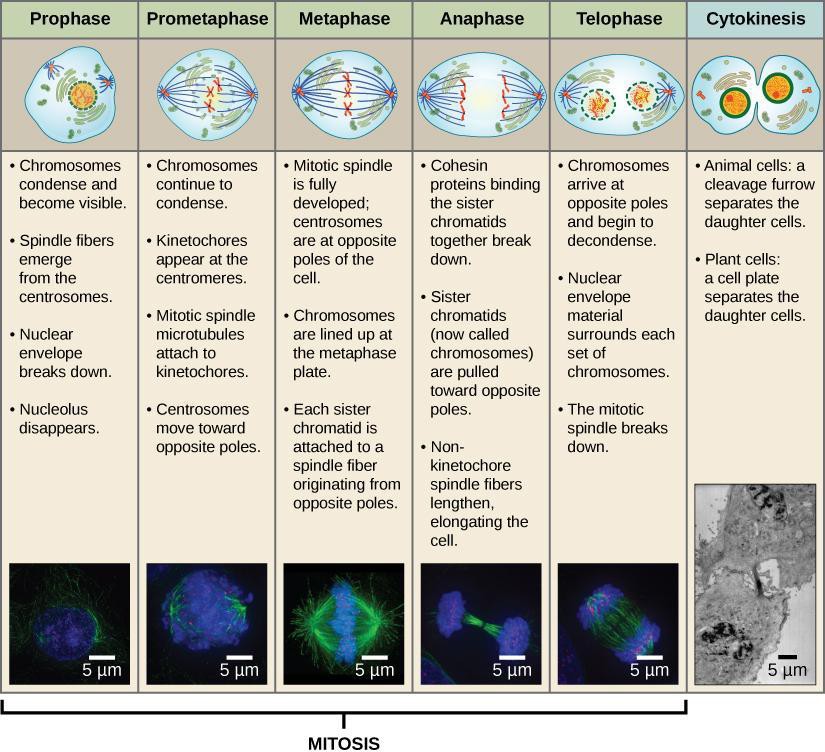
During prophase, the chromosomes coil up, and sister chromatids become visible under a microscope. The nuclear membrane surrounding the chromosomes also disappears. In metaphase, the sister chromatids align in the center of the cell, attached to spindle fibers. During anaphase, the sister chromatids separate and move to opposite poles of the cell. In telophase, the chromosomes arrive at the poles and begin to decondense while the nucleus reforms. Figure 13.3 makes it look like the phases are very distinct. The phases, however, transition without stopping. Notice also other important structures, such as the spindle fibers and kinetochores on the centromeres of each chromosome. Each pair of sister chromatids has a protein structure, called a kinetochore, which becomes attached to spindle fibers. The spindle fibers pull the sister chromatids apart, toward opposite ends of the cell.
Cancer cells are not healthy cells. Healthy cells die through programmed cell death or apoptosis after a number of generations of cell divisions. Cells become mature or differentiate, which enables them to carry out their function in the body. Cancer is the uncontrolled growth and mitotic division of cells which can be caused by mutations in genes. Some chemotherapy drugs, including taxanes such as Taxol from the Yew tree and alkaloids from the Vinca plant, interfere with mitosis by binding to microtubules and preventing spindle fibers from separating sister chromatids, thus leading to cell death.
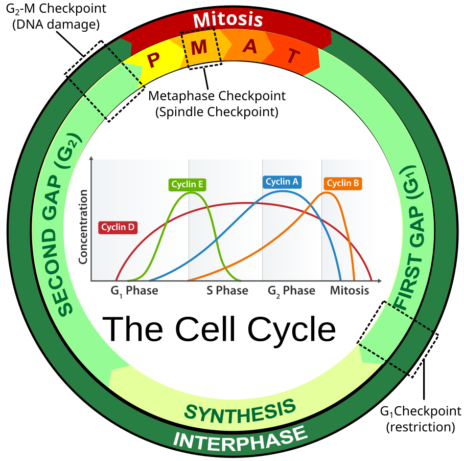 Figure 13.1: Eukaryotic cell cycle is governed by expression of cyclin proteins along with their activity. Credit: Jeremy Seto (CC-BY-SA)
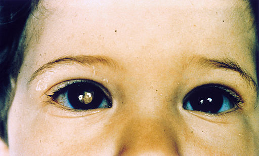 https://commons.wikimedia.org/wiki/File:Retinoblastoma.jpg Figure 13.2: The checkpoints of the Cell Cycle are prone to DNA damage which can cause a disease like cancer. Exposure of tumor in the right eye by white light reflection.
Defective Cell Cycle Checkpoints: A white light shone on a child’s eye should yield a clear view of the retina. In the above image, the right eye shows a white light reflecting and indicates a retinoblastoma (Figure 13.2). This cancer is caused by a defect in the Rb gene, a tumor suppressor gene. This defect permits the continuation of the cell cycle despite damage to DNA. Retinoblastoma is the most common primary childhood cancer which often stems from a genetic background.
Safety Precautions
- Be careful handling glass slides, as the edges may be sharp.
- Observe proper use of the microscope; avoid handling the electric cord with wet hands.
- Do not use the coarse adjustment knob of the microscope at higher magnifications.
- There is a separate marked disposal for sharp objects like broken glass. If you cannot locate it, inform your Instructor/Lab Technician immediately of any broken glassware, as it could cause serious injuries.
For this activity, you will need the following:
-
-
- Prepared slide of whitefish blastula cells, or use online images and resources
-
For this activity, you will work in pairs.
Structured Inquiry
Step 1: Using Figure 13.1, predict the percent of time that a cell would spend in each phase. Based on this prediction, how much time do you think cells will spend in interphase as opposed to mitosis? Write your prediction on your lab paper.
Step 2: Look at Figure 13.3. On your paper, make a table defining the characteristics of the stages of mitosis, as well as interphase, that you can use to identify each stage under the microscope. Note: There are four major phases of mitosis, plus prometaphase, which is a transition phase between prophase and metaphase.
Step 3: Using the prepared slide, record the number of cells in each phase of the cell cycle. Use the method shown in Figure 13.4 to count the cells. Record your count in a data table like that shown in Table 13.1. Share your data with the class to create a group total count.
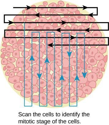
Table 13.1: Results of Cell Stage Identification
|
Phase or Stage |
Individual Totals |
Class or Group Totals |
Percent |
|---|---|---|---|
|
Interphase |
|
|
|
|
Prophase |
|
|
|
|
Metaphase |
|
|
|
|
Anaphase |
|
|
|
|
Telophase |
|
|
|
|
Cytokinesis |
|
|
|
|
Totals |
|
|
100% |
Step 4: In your data table, calculate the percentage of cells in each phase.
Step 5: Are the predictions you made supported by your data (observations and calculations)? Do your results match the diagram as presented in Figure 13.1? Why or why not?
Assessments
- Explain why interphase could be the longest phase and mitosis and cytokinesis are generally much shorter phases of the cell cycle.
- Explain the importance of spindle fibers in mitosis and why antimitotic drugs that block spindle fiber formation are used to treat cancer.
Activity
- In a plant, where would you likely find cells that are actively undergoing mitosis? Where would you likely find cells that aren’t actively undergoing mitosis? Explain your answers.
- Think about how a plant grows. In which parts of the plant would many cells likely be undergoing mitosis?
- Discuss the answers to the questions with a partner (think, pair, share) and then the class.
Observe the Stages of Mitosis in Onion Root Cells
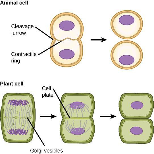
Plant cells also use mitosis for growth, maintenance, and repair. The plant’s cell wall, as well as the nuclear material, makes observing mitosis much easier. Figure 13.5 shows some of the similarities and differences between plant and animal mitosis. For example, during cytokinesis, an animal cell pinches apart into two daughter cells, while a plant cell develops a new cell wall, called a cell plate, between the new daughter cells.
Safety Precautions
- Be careful handling glass slides, as the edges may be sharp.
- Observe proper use of the microscope; avoid handling the electric cord with wet hands.
- Do not use the coarse adjustment knob of the microscope at higher magnifications.
- Inform your teacher immediately of any broken glassware, as it could cause injuries.
For this activity, you will need the following:
- Prepared microscope slides of stained onion root tips or online images and resources

https://commons.wikimedia.org/wiki/File:Mitosis_(261_15)_Pressed;_root_meristem_of_onion.jpg Stained actively dividing cells of onion root tip Figure 13.5a
This video provides a demo of materials including onion or garlic bulbs, germination of root, preparation of root tip, staining, and observing under the microscope:
For this activity, you will work in pairs.
Structured Inquiry
Step 1: Hypothesize what regions of the onion root tip might differ in the number of cells undergoing mitosis. What is your reasoning behind this hypothesis? Record your answers on your paper.
Step 2: Make a drawing of each phase of mitosis, as well as interphase, in a plant cell. Label, in each drawing, the defining features that you will look for when identifying each stage under the microscope.
Step 3: Are the predictions you made in Step 1 supported by your observations? Why do you think cells undergoing mitosis in the onion root cell are distributed the way they are? What differences between plant and animal cells are visible under the microscope slide?
Assessments
- Describe some similarities and some differences between animal and plant mitosis.
- Based on your observations, explain how onion roots grow at the cellular level.
Activity
- Looking at Figures 13.3 and 13.6, compare the outcomes of mitosis versus meiosis.
- After meiosis, some daughter cells may not contain the correct number of chromosomes. What failure in the meiosis process could cause this to occur?
- Discuss the answers to questions with a partner (think, pair, share) and then the class.
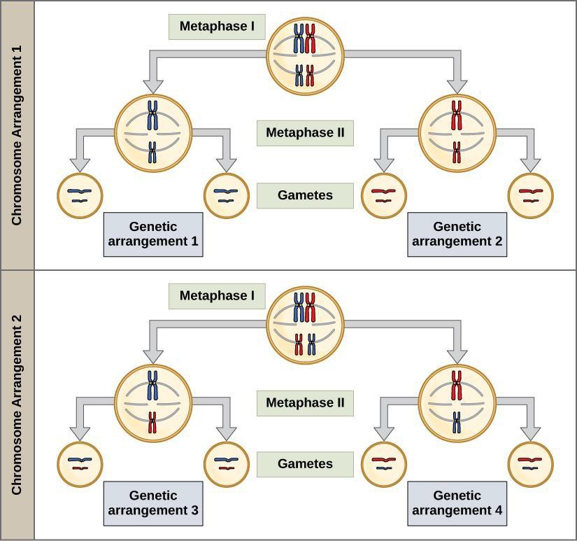
Figure 13.6: Overview of meiosis typically in sexually reproducing organisms that reduces the number of chromosomes to produce gametes or sex cells (egg in female and sperm in male)
Meiosis and Fertilization
Meiosis is the process in sexually reproducing eukaryotes that forms sex cells or gametes, which include sperm and eggs (ova). To avoid doubling the number of chromosomes in each generation, reduction division (halving the number of chromosomes) in gamete production is necessary. Chromosomes are typically diploid (2N) or occur as double sets (homologous pairs) in each nucleus. Each homolog of a pair has the same sites or loci for the same genes. You might recognize that you have one set of chromosomes from your mother and the remaining set from your father. Meiosis reduces the number of chromosomes to a haploid (1N) or single set. Meiosis uses very similar mechanisms to mitosis. There are, however, several significant differences. The source cells for meiosis are found in the reproductive organs of animals (gonads: ovaries; oogenesis, where the eggs are produced and testes; spermatogenesis, where the sperms are produced) and plants (flowers: ovaries and anthers). Major differences between mitosis and meiosis include the association of homologous chromosomes (sister chromatids of the same chromosomes) attached as a tetrad group of four, as well as crossover of chromosome regions during prophase I of meiosis I. Meiosis shuffles the genetic material so that each resulting cell carries a new and unique set of genes in a process of independent assortment. Crossover provides one source of genetic variation in offspring.
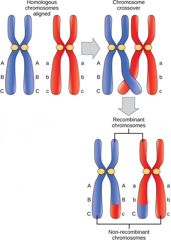
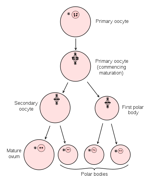
Figure 13.8: Oogenesis: In the human female genitalia reproductive structures, a growth process in which the primary egg cell (or ovum) matures into an ovum (1N)
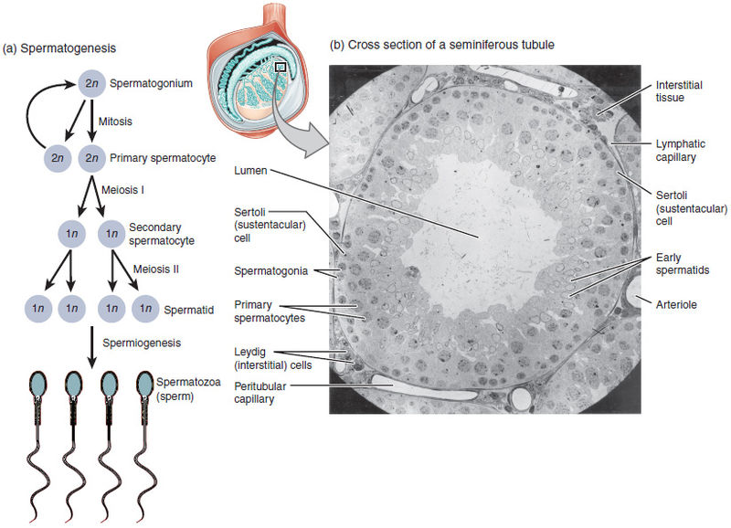
Figure 13.9: Spermatogenesis: In this process, haploid spermatozoa (1N) develop from germ cells in the seminiferous tubules of the testis male genitalia reproductive structures
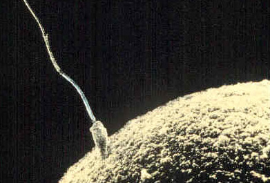
Figure 13.10: Fertilization: The union or fusion of the 2 gametes, ovum (1N) haploid and spermatozoa (1N) haploid in which the fertilized egg or zygote (2N) diploid has the double chromosome set restored. The size of the egg is emphasized by the sperm in contact
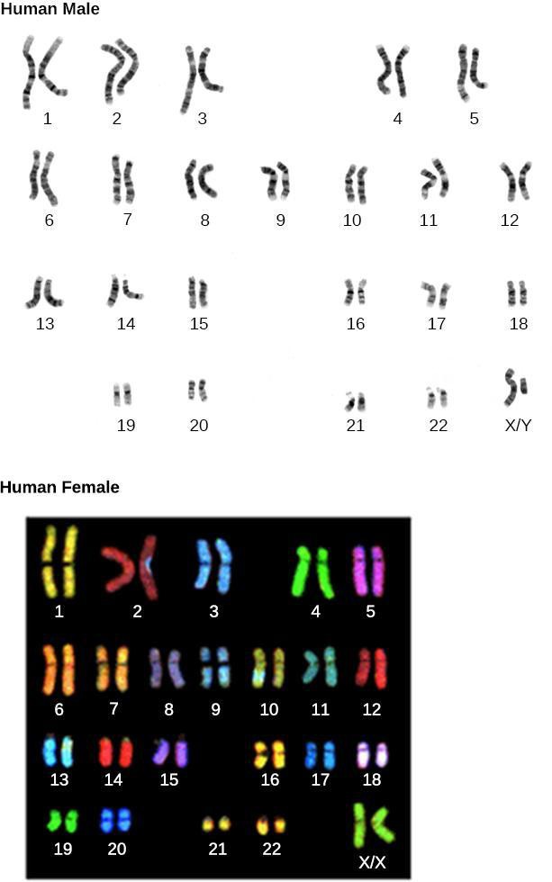
Humans have 23 different chromosomes. As stated, we have a diploid (2N) set of chromosomes: a full set of 23 chromosomes received from the sperm and another set of 23 from the ovum (egg) during fertilization. See Figure 13.11. During metaphase I of meiosis I, homologous chromosomes are separated. The chromosome number is reduced from diploid (2N) to haploid (1N) by the end of meiosis I, with each cell retaining duplicate sister chromatids. Meiosis II results in four haploid (1N) cells in sperm, each with only one set of each of the 23 chromosomes. In female humans, meiosis results in uneven division of the cytoplasm and the formation of two polar bodies (nuclei only) that are not involved in fertilization. One note about human fertilization: meiosis II is completed only after the sperm has entered the ovum.
There are several malfunctions that can occur during meiosis. Nondisjunction occurs when the sister chromatids in tetrads separate unevenly. Nondisjunction results in one cell getting an extra chromosome while another cell is missing a chromosome. Nondisjunction is associated with certain human genetic disorders. For example, Down syndrome is usually caused by nondisjunction that results in three copies of chromosome 21. Translocations can occur when chromosomes exchange genetic information with nonhomologous chromosomes. Here is a a more detailed list.
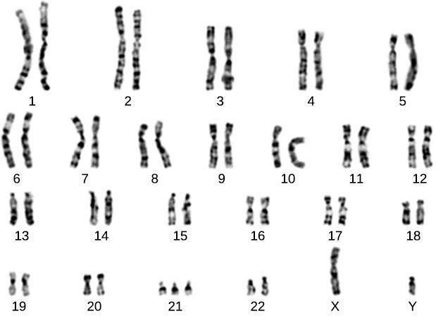
Karyotypes (chromosome spreads) are made by stopping cells in mitosis with a chemical and then dyeing with Giemsa stain. A picture is taken through a microscope and then digitally enlarged to see the chromosomal banding, or G-bands. Dark and light banding patterns help identify chromosomes and alterations to normal chromosomes. The chromosome spreads can be seen in Figure 13.11. Human females have two X chromosomes, and males have one X and one Y.
Safety Precautions
None
For this activity, you will need the following:
- Images of karyotypes
For this activity, you will work in pairs.
Structured Inquiry
Step 1: A karyotype shows the number and appearance of chromosomes in the nucleus of a cell. Predict what you would look for in an abnormal karyotype. Record your predictions on your paper.
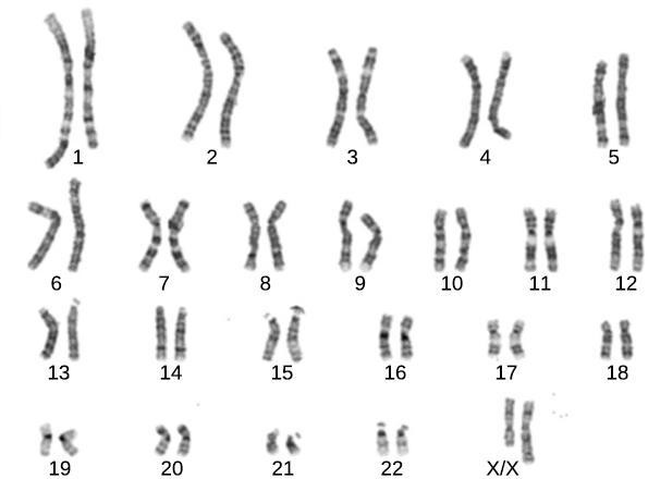
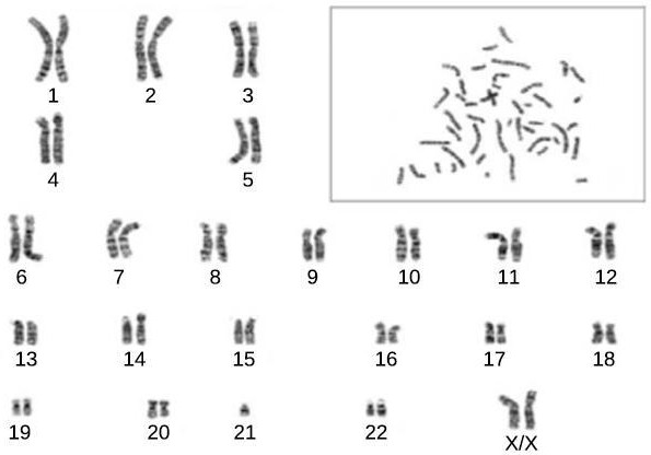
Step 2: Examine the three karyotypes 1, 2, and 3 shown in Figure 13.12, Figure 13.13, and Figure 13.14 respectively (below). Compare these karyotypes to the normal karyotypes shown in Figure 13.11 (above). Can you tell if the individual is female, male, or indeterminate (does not have a normal distribution of sex chromosomes)? Record any abnormalities on your paper, and research the meaning of the changes in chromosome number or appearance.
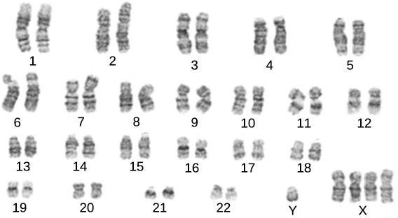
Step 3: Critical Analysis: What are the implications of nondisjunction in a karyotype? Using Karyotype 1 (Figure 13.12) as an example, explain the implications of the nondisjunction malfunction during meiosis for the chromosome count of the daughter cells.
Assessments
- This Robertsonian translocation involves a fusion of the long arms of two non-homologous chromosomes during meiosis. A patient possesses a Robertsonian translocation among chromosome 14 onto chromosome 13. No genetic material is missing from either chromosome. Explain why it is possible that there is no abnormality in the person but could be in the patient’s offspring.
 chromosomes.
 - Crossing over of chromosomes during prophase I is common. How does this process increase the genetic diversity of the daughter cells?
identical copies of the same chromosome
stage of mitosis during which chromosomes condense and the mitotic spindle begins to form
stage of mitosis where the chromosomes are at their most compact
stage of mitosis during which sister chromatids are separated from each other
stage of mitosis during which chromosomes arrive at opposite poles, decondense, and are surrounded by a new nuclear envelope
protein structure associated with the centromere of each sister chromatid that attracts and binds spindle microtubules during prometaphase
structure formed during plant cell cytokinesis by Golgi vesicles, forming a temporary structure (phragmoplast) and fusing at the metaphase plate; ultimately leads to the formation of cell walls that separate the two daughter cells
halving the number of chromosomes
sister chromatids of the same chromosomes
two duplicated homologous chromosomes (four chromatids) bound together by chiasmata during prophase I
cell with two sets of chromosomes
union of two haploid cells from two individual organisms
failure of synapsed homologs to completely separate and migrate to separate poles during the meiosis’s first cell division
process by which one chromosome segment dissociates and reattaches to a different, nonhomologous chromosome
a cell’s chromosomes
