11 DNA EXTRACTION
Learning Objectives
- Extract DNA from the cells of strawberry fruit tissue
- Demonstrate DNA replication process with modeling
- Demonstrate transcription and translation using a specific sequence
Activity
- In a plant like the strawberry, where would you likely find DNA?
- Discuss the answers to the questions with a partner (think, pair, share) and then your group.
DNA Extraction from Strawberries
The first step in working with nucleic acids (DNA and RNA) is to remove the molecules from inside the cell. Different types of cells need to be processed differently in order to release nucleic acids. All cells have a cell membrane, a phospholipid bilayer that separates the internal environment of the cell from the external environment. In eukaryotes, DNA is housed inside the nucleus of the cell, which is surrounded by the nuclear membrane, a second double-layered membrane, also composed largely of lipid molecules. When extracting DNA from plant cells, the cell wall must also be considered; some types of plant tissue require grinding or flash-freezing in order to break through the tough cell wall.
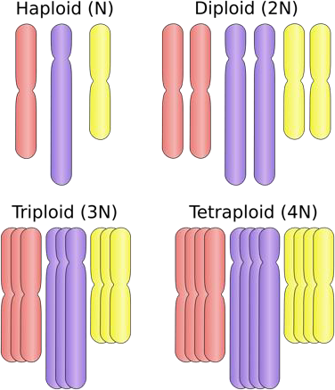
Strawberry fruit tissue is an excellent type of tissue to use for demonstration of DNA extraction. First, ripe strawberries are soft and juicy; as the fruit matures, the cells fill up with water and sugar, which make the fruit so delicious! Second, as the strawberry ripens, a series of chemical reactions take place within the cells that lead to the breakdown of long-chain polysaccharides, like cellulose and pectin, that make the cell wall tough. Lastly, cultivated strawberries (Fragaria x ananassa) are the product of a hybridization between two other strawberry species, and they have an octoploid genome, meaning they have eight sets of chromosomes inside their cells. That translates to lots of molecules of DNA, which increases our yield and makes the DNA easier to visualize.
This protocol for extraction of DNA is based largely on the principle of solubility. Solubility refers to the ability of one substance (the solute) to dissolve in another substance (the solvent). Recall that polar substances dissolve easily in polar solvents, but do not dissolve easily in nonpolar solvents, a phenomenon commonly referred to as “like dissolves like.” Water is a polar solvent, and molecules that dissolve easily in water are referred to as hydrophilic. DNA molecules are hydrophilic because the sugar-phosphate backbone of the molecules are highly polar. This means that DNA dissolves in water, so in this experiment, the DNA that is released when the cells are crushed dissolves in the juice/extraction buffer mixture.
Remember, there are two key ingredients in the DNA extraction buffer aside from the water: dish soap and salt. The dish soap acts to break up the lipid molecules that form the cell membrane and the nuclear membrane, which lyses the cell and releases the cellular contents, including DNA. The salt has two functions in the extraction process. It helps to neutralize the charge on the sugar-phosphate backbone, making DNA less soluble in water, and it also makes the DNA molecules stick to each other, so they are easier to visualize and remove from the solution.
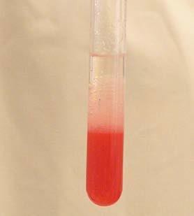
Although the chemical reactions described above are all happening when you add the buffer and crush the strawberries, they are not visible to the naked eye. However, the addition of the cold ethanol caused a much more dramatic result! Ethanol is a nonpolar solvent, and when it is added to the solution of strawberry juice and extraction buffer, the DNA precipitates out of the solution. A precipitation reaction is a chemical reaction that causes a solid substance to emerge from a liquid solution. In this experiment, the addition of ethanol to the reaction forces DNA to precipitate out of solution, which we can then spool onto the wooden skewer.
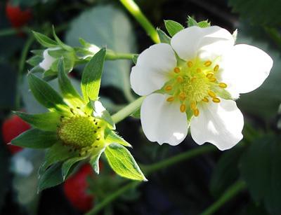
Strawberry flowers; picture from Wikimedia Commons
Illustrations of ploidy levels; N=number of chromosomes in one set. Strawberries (Fragaria x ananassa) are octoploid (8N); illustration from Wikimedia Commons
Safety Precautions
For this activity, you will need the following:
- Plastic zipper bag
- 1 fresh strawberry
- 10 mL DNA extraction buffer
- Gauze squares
- Funnel
- Ice cold ethanol
- Plastic transfer pipettes
- Clear glass test tube
- Wooden skewer
- Microcentrifuge tubes
To make DNA extraction buffer (optional), combine the following:
- 45 mL DI water
- 5mL liquid dish soap
- 0.75 g NaCl (table salt)
For this activity, you will work in pairs.
Structured Inquiry
Step 1: Place strawberry into plastic zipper bag and add 10mL of DNA extraction buffer. Seal the bag tightly.
Gently, but thoroughly crush the strawberry inside the bag for about one minute.
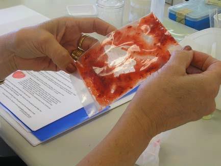
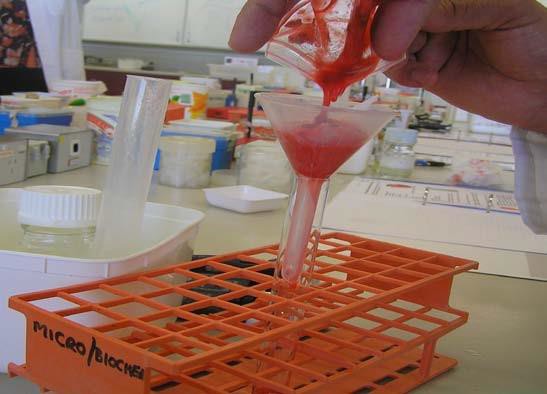
Line the funnel with a gauze square. Place the funnel into the test tube.
Pour the strawberry juice/DNA extraction buffer mixture into the funnel so the juice passes through the gauze and into the test tube. Use the gauze to strain the strawberry mixture so that only the juice flows into the tube and the pulp is retained in the gauze.
Discard the gauze and the strawberry pulp.
Step 2: After straining the strawberry juice/DNA extraction buffer mix, do you anticipate being able to see DNA with the naked eye? What does DNA look like? Write your hypotheses and expectations on your lab paper.
Layer an equal volume of ice cold ethanol on top of the strawberry juice mixture in the test tube using the plastic transfer pipette.
Step 3: Hold the tube still at eye level (DO NOT shake) and observe what happens at the interface of the alcohol and the strawberry juice when you twirl your wooden skewer through the interface of the solutions.
Use your wooden skewer to transfer your strawberry DNA into a microcentrifuge tube. Add a small amount of ethanol to the tube to prevent your DNA from drying out.
Step 4: Critical Analysis: You now have a clump of aggregated DNA molecules that you can see! This DNA contains all the genes within the strawberry genome.
Assessment
- Given what you know about DNA, what could the DNA be used for after it has been extracted?
- Discuss with your partner.
References: Jones, Carolyn. DNA Extraction from Strawberries. https://www.murdoch.edu.au/Biotech-out-of-the-box/_document/Kit-Handout-Sheets/DNA-extraction-from-strawberries.pdfOpenStax CNX. 2018. https://cnx.org/contents/k6E8Y6SA@8/Primary-DNA-Molecular-Structure University of Missouri, Division of Plant Sciences. 2018. Strawberry: A Brief History. https://ipm.missouri.edu/meg/2012/5/Strawberry-A-Brief-History/
DNA REPLICATION
In the 1950s, Francis Crick and James Watson worked together to determine the structure of DNA at the University of Cambridge, England. Other scientists like Linus Pauling and Maurice Wilkins were also actively exploring this field. Pauling previously had discovered the secondary structure of proteins using X-ray crystallography. In Wilkins’ lab, researcher Rosalind Franklin was using X-ray diffraction methods to understand the structure of DNA. Watson and Crick were able to piece together the puzzle of the DNA molecule on the basis of Franklin’s data because Crick had also studied X-ray diffraction (Figure 14.6). In 1962, James Watson, Francis Crick, and Maurice Wilkins were awarded the Nobel Prize in Medicine. Unfortunately, by then Franklin had died, and Nobel prizes are not awarded posthumously.
Figure 14.6 The X-ray diffraction pattern of DNA, which helped to elucidate its double-helix structure.
Watson and Crick proposed that DNA is made up of two strands that are twisted around each other to form a right-handed helix. Base pairing takes place between a purine and pyrimidine on opposite strands, so that A pairs with T, and G pairs with C (suggested by Chargaff’s Rules). Thus, adenine and thymine are complementary base pairs, and cytosine and guanine are also complementary base pairs. The base pairs are stabilized by hydrogen bonds: adenine and thymine form two hydrogen bonds and cytosine and guanine form three hydrogen bonds. The two strands are anti-parallel in nature; that is, the 3′ end of one strand faces the 5′ end of the other strand. The sugar and phosphate of the nucleotides form the backbone of the structure, whereas the nitrogenous bases are stacked inside, like the rungs of a ladder. Each base pair is separated from the next base pair by a distance of 0.34 nm, and each turn of the helix measures 3.4 nm. Therefore, 10 base pairs are present per turn of the helix. The diameter of the DNA double-helix is 2 nm, and it is uniform throughout. Only the pairing between a purine and pyrimidine and the antiparallel orientation of the two DNA strands can explain the uniform diameter. The twisting of the two strands around each other results in the formation of uniformly spaced major and minor grooves (Figure 14.7).
Figure 14.7 DNA has (a) a double helix structure and (b) phosphodiester bonds; the dotted lines between Thymine and Adenine and Guanine and Cytosine represent hydrogen bonds. The (c) major and minor grooves are binding sites for DNA binding proteins during processes such as transcription (the copying of RNA from DNA) and replication.
DNA Sequencing Techniques
Until the 1990s, the sequencing of DNA (reading the sequence of DNA) was a relatively expensive and long process. Using radiolabeled nucleotides also compounded the problem through safety concerns. With currently available technology and automated machines, the process is cheaper, safer, and can be completed in a matter of hours. Fred Sanger developed the sequencing method used for the human genome sequencing project, which is widely used today (Figure 14.8).
Assessment
- What is the central dogma of biology?
- Describe how the result of replication differs from the result of transcription.
- Are transcription and translation happening in your cells right now?
the semipermeable membrane surrounding the cytoplasm of a cell
a two-layered arrangement of phosphate and lipid molecules that form a cell membrane, the hydrophobic lipid ends facing inward and the hydrophilic phosphate ends facing outward
is made up of two lipid bilayer membranes that in eukaryotic cells surround the nucleus, which encloses the genetic material. The nuclear envelope consists of two lipid bilayer membranes: an inner nuclear membrane and an outer nuclear membrane
rigid cell covering comprised of various molecules that protects the cell, provides structural support, and gives shape to the cell
a method to purify DNA by using physical and/or chemical methods from a sample separating DNA from cell membranes, proteins, and other cellular components
the ability to be dissolved, especially in water
substance that resists a change in pH by absorbing or releasing hydrogen or hydroxide ions
a buffer solution used for the purpose of breaking open cells for use in molecular biology experiments that analyze the compounds of the cells
